
автордың кітабын онлайн тегін оқу A Treatise on Anatomy, Physiology, and Hygiene (Revised Edition)
The Project Gutenberg EBook of A Treatise on Anatomy, Physiology, and
Hygiene (Revised Edition), by Calvin Cutter
This eBook is for the use of anyone anywhere at no cost and with
almost no restrictions whatsoever. You may copy it, give it away or
re-use it under the terms of the Project Gutenberg License included
with this eBook or online at www.gutenberg.net
Title: A Treatise on Anatomy, Physiology, and Hygiene (Revised Edition)
Author: Calvin Cutter
Release Date: November 24, 2009 [EBook #30541]
Language: English
*** START OF THIS PROJECT GUTENBERG EBOOK TREATISE ON ANATOMY (REVISED) ***
Produced by Bryan Ness, Dan Horwood and the Online
Distributed Proofreading Team at http://www.pgdp.net (This
file was produced from images generously made available
by The Internet Archive/American Libraries.)
A
TREATISE
ON
ANATOMY, PHYSIOLOGY,
AND HYGIENE
DESIGNED
FORCOLLEGES, ACADEMIES, AND FAMILIES.
BY CALVIN CUTTER, M.D.
WITH ONE HUNDRED AND FIFTY ENGRAVINGS.
REVISED STEREOTYPE EDITION.
NEW YORK:
CLARK, AUSTIN AND SMITH.
CINCINNATI:—W. B. SMITH & CO.
ST. LOUIS, MO.:—KEITH & WOODS.
1858.
Entered according to Act of Congress, in the year 1852, by
CALVIN CUTTER, M. D.,
In the Clerk’s Office of the District Court of the District of
Massachusetts
.C. A. ALVORD,
Printer,
No. 15 Vandewater Street, N.
Y.
PREFACE.
Agesilaus, king of Sparta, when asked what things boys should learn, replied, “Those which they will practise when they become men.” As health requires the observance of the laws inherent to the different organs of the human system, so not only boys, but girls, should acquire a knowledge of the laws of their organization. If sound morality depends upon the inculcation of correct principles in youth, equally so does a sound physical system depend on a correct physical education during the same period of life. If the teacher and parents who are deficient in moral feelings and sentiments, are unfit to communicate to children and youth those high moral principles demanded by the nature of man, so are they equally incompetent directors of the physical training of the youthful system, if ignorant of the organic laws and the physiological conditions upon which health and disease depend.
For these reasons, the study of the structure of the human system, and the laws of the different organs, are subjects of interest to all,—the young and the old, the learned and the unlearned, the rich and the poor. Every scholar, and particularly every young miss, after acquiring a knowledge of the primary branches,—as spelling, reading, writing, and arithmetic,—should learn the structure of the human system, and the conditions upon which health and disease depend, as this knowledge will be required in practice in after life.
“It is somewhat unaccountable,” says Dr. Dick, “and not a little inconsistent, that while we direct the young to look abroad over the surface of the earth, and survey its mountains, rivers, seas, and continents, and guide their views to the regions of the firmament, where they may contemplate the moons of Jupiter, the rings of Saturn, and thousands of luminaries placed at immeasurable distances, ... that we should never teach them to look into themselves; to consider their own corporeal structures, the numerous parts of which they are composed, the admirable functions they perform, the wisdom and goodness displayed in their mechanism, and the lessons of practical instruction which may be derived from such contemplations.”
Again he says, “One great practical end which should always be kept in view in the study of physiology, is the invigoration and improvement of the corporeal powers and functions, the preservation of health, and the prevention of disease.”
The design of the following pages is, to diffuse in the community, especially among the youth, a knowledge of Human Anatomy, Physiology, and Hygiene. To make the work clear and practical, the following method has been adopted:—
1st. The structure of the different organs of the system has been described in a clear and concise manner. To render this description more intelligible, one hundred and fifty engravings have been introduced, to show the situation of the various organs. Hence the work may be regarded as an elementary treatise on anatomy.
2d. The functions, or uses of the several parts have been briefly and plainly detailed; making a primary treatise on human physiology.
3d. To make a knowledge of the structure and functions of the different organs practical, the laws of the several parts, and the conditions on which health depends, have been clearly and succinctly explained. Hence it may be called a treatise on the principles of hygiene, or health.
To render this department more complete, there has been added the appropriate treatment for burns, wounds, hemorrhage from divided arteries, the management of persons asphyxiated from drowning, carbonic acid, or strangling, directions for nurses, watchers, and the removal of disease, together with an Appendix, containing antidotes for poisons, so that persons may know what should be done, and what should not be done, until a surgeon or physician can be called.
In attempting to effect this in a brief elementary treatise designed for schools and families, it has not been deemed necessary to use vulgar phrases for the purpose of being understood. The appropriate scientific term should be applied to each organ. No more effort is required to learn the meaning of a proper, than an improper term. For example: a child will pronounce the word as readily, and obtain as correct an idea, if you say lungs, as if you used the word lights. A little effort on the part of teachers and parents, would diminish the number of vulgar terms and phrases, and, consequently, improve the language of our country. To obviate all objections to the use of proper scientific terms, a Glossary has been appended to the work.
The author makes no pretensions to new discoveries in physiological science. In preparing the anatomical department, the able treatises of Wilson, Cruveilhier, and others have been freely consulted. In the physiological part, the splendid works of Carpenter, Dunglison, Liebig, and others have been perused. In the department of hygiene many valuable hints have been obtained from the meritorious works of Combe, Rivers, and others.
We are under obligations to R. D. Mussey, M. D., formerly Professor of Anatomy and Surgery, Dartmouth College, N. H., now Professor of Surgery in the Ohio Medical College; to J. E. M’Girr, A. M., M. D., Professor of Anatomy, Physiology, and Chemistry, St. Mary’s University, Ill.; to E. Hitchcock, Jr., A. M., M. D., Teacher of Chemistry and Natural History, Williston Seminary, Mass.; to Rev. E. Hitchcock, D. D., President of Amherst College, Mass., who examined the revised edition of this work, and whose valuable suggestions rendered important aid in preparing the manuscript for the present stereotype edition.
We return our acknowledgments for the aid afforded by the Principals of the several Academies and Normal Schools who formed classes in their institutions, and examined the revised edition as their pupils progressed, thus giving the work the best possible test trial, namely, the recitation-room.
To the examination of an intelligent public, the work is respectfully submitted by
CALVIN CUTTER.
Warren, Mass., Sept. 1, 1852.
TO TEACHERS AND PARENTS.
As the work is divided into chapters, the subjects of which are complete in themselves, the pupil may commence the study of the structure, use, and laws of the several parts of which the human system is composed, by selecting such chapters as fancy or utility may dictate, without reference to their present arrangement,—as well commence with the chapter on the digestive organs as on the bones.
The acquisition of a correct pronunciation of the technical words is of great importance, both in recitation and in conversation. In this work, the technical words interspersed with the text, have been divided into syllables, and the accented syllables designated. An ample Glossary of technical terms has also been appended to the work, to which reference should be made.
It is recommended that the subject be examined in the form of topics. The questions in Italics are designed for this method of recitation. The teacher may call on a pupil of the class to describe the anatomy of an organ from an anatomical outline plate; afterwards call upon another to give the physiology of the part, while a third may state the hygiene, after which, the questions at the bottom of the page may be asked promiscuously, and thus the detailed knowledge of the subject possessed by the pupils will be tested.
At the close of the chapters upon the Hygiene of the several portions of the system, it is advised that the instructor give a lecture reviewing the anatomy, physiology, and hygiene, of the topic last considered. This may be followed by a general examination of the class upon the same subject. By this course a clear and definite knowledge of the mutual relation of the Anatomy, Physiology, and Hygiene, of different parts of the human body, will be presented.
We also suggest the utility of the pupils’ giving analogous illustrations, examples, and observations, where these are interspersed in the different chapters, not only to induce inventive thought, but to discipline the mind.
To parents and others we beg leave to say, that about two thirds of the present work is devoted to a concise and practical description of the uses of the important organs of the human body, and to show how such information may be usefully applied, both in the preservation of health, and the improvement of physical education. To this have been added directions for the treatment of those accidents which are daily occurring in the community, making it a treatise proper and profitable for the FAMILY LIBRARY, as well as the school-room.
CONTENTS.
Chapter.
Page.
1.
General Remarks,
132.
Structure of Man,
173.
Chemistry of the Human Body,
254.
Anatomy of the Bones,
295.
Anatomy of the Bones, continued,
396.
Physiology of the Bones,
487.
Hygiene of the Bones,
538.
Anatomy of the Muscles,
649.
Physiology of the muscles,
7610.
Hygiene of the Muscles,
8511.
Hygiene of the Muscles, continued,
9612.
Anatomy of the Teeth,
10512.
Physiology of the Teeth,
10912.
Hygiene of the Teeth,
11013.
Anatomy of the Digestive Organs,
11314.
Physiology of the Digestive Organs,
12415.
Hygiene of the Digestive Organs,
12916.
Hygiene of the Digestive Organs, continued,
14217.
Anatomy of the Circulatory Organs,
15418.
Physiology of the Circulatory Organs,
16419.
Hygiene of the Circulatory Organs,
17220.
Anatomy of the Lymphatic Vessels,
18120.
Physiology of the Lymphatic Vessels,
18320.
Hygiene of the Lymphatic Vessels,
18821.
Anatomy of the Secretory Organs.
19221.
Physiology of the Secretory Organs,
19321.
Hygiene of the Secretory Organs,
19722.
Nutrition,
20022.
Hygiene of Nutrition,
20523.
Anatomy of the Respiratory Organs,
20924.
Physiology of the Respiratory Organs,
21725.
Hygiene of the Respiratory Organs,
22826.
Hygiene of the Respiratory Organs, continued,
23927.
Animal Heat,
25228.
Hygiene of Animal Heat,
26129.
Anatomy of the Vocal Organs,
26829.
Physiology of the Vocal Organs,
27230.
Hygiene of the Vocal Organs,
27431.
Anatomy of the Skin,
28232.
Physiology of the Skin,
29333.
Hygiene of the Skin,
30134.
Hygiene of the Skin, continued,
31135.
Appendages of the Skin,
32236.
Anatomy of the Nervous System,
32737.
Anatomy of the Nervous System, continued,
34038.
Physiology of the Nervous System,
34639.
Hygiene of the Nervous System,
35840.
Hygiene of the Nervous System, continued,
36841.
The Sense of Touch,
37842.
Anatomy of the Organs of Taste,
38442.
Physiology of the Organs of Taste,
38643.
Anatomy of the Organs of Smell,
38943.
Physiology of the Organs of Smell,
39144.
Anatomy of the Organs of Vision,
39445.
Physiology of the Organs of Vision,
40445.
Hygiene of the Organs of Vision,
41046.
Anatomy of the Organs of Hearing,
41447.
Physiology of the Organs of Hearing,
42047.
Hygiene of the Organs of Hearing,
42248.
Means of preserving the Health,
42549.
Directions for Nurses,
432APPENDIX,
439GLOSSARY,
451INDEX,
463ANATOMY, &c.
CHAPTER I.
GENERAL REMARKS.
1. Anatomy is the science which treats of the structure and relations of the different parts of animals and plants.
2. It is divided into Vegetable and Animal anatomy. The latter of these divisions is subdivided into Human anatomy, which considers, exclusively, human beings; and Comparative anatomy, which treats of the mechanism of the lower orders of animals.
3. Physiology treats of the functions, or uses of the organs of animals and plants. Another definition is, “the science of life.”
4. This is also divided into Vegetable and Animal physiology, as it treats of the vegetable or animal kingdom; and into Human and Comparative physiology, as it describes the vital functions of man or the inferior animals.
5. Hygiene is the art or science of maintaining health, or a knowledge of those laws by which health may be preserved.
6. The kingdom of nature is divided into organic and inorganic bodies. Organic bodies possess organs, on whose action depend their growth and perfection. This division includes animals and plants. Inorganic bodies are devoid of organs, or instruments of life. In this division are classed the earths, metals, and other minerals.
1. What is anatomy? 2. How is it divided? How is the latter division subdivided? 3. What is physiology? Give another definition. 4. How is physiology divided? Give a subdivision. 5. What is hygiene? 6. Define organic bodies.
7. In general, organic matter differs so materially from inorganic, that the one can readily be distinguished from the other. In the organic world, every individual of necessity springs from some parent, or immediate producing agent; for while inorganic substances are formed by chemical laws alone, we see no case of an animal or plant coming into existence by accident or chance, or chemical operations.
8. Animals and plants are supported by means of nourishment, and die without it. They also increase in size by the addition of new particles of matter to all parts of their substances; while rocks and minerals grow only by additions to their surfaces.
9. “Organized bodies always present a combination of both solids and fluids;—of solids, differing in character and properties, arranged into organs, and these endowed with functional powers, and so associated as to form of the whole a single system;—and of fluids, contained in these organs, and holding such relation to the solids that the existence, nature, and properties of both mutually and necessarily depend on each other.”
10. Another characteristic is, that organic substances have a certain order of parts. For example, plants possess organs to gain nourishment from the soil and atmosphere, and the power to give strength and increase to all their parts. And animals need not only a digesting and circulating apparatus, but organs for breathing, a nervous system, &c.
6. Define inorganic bodies. 7. What is said of the difference, in general, between organic and inorganic bodies? 8. What of the growth of organic and inorganic bodies? 9. What do organized bodies always present? 10. Give another characteristic of organized substances.
11. Individuality is an important characteristic. For instance, a large rock may be broken into a number of smaller pieces, and yet every fragment will be rock; but if an organic substance be separated into two or more divisions, neither of them can be considered an individual. Closely associated with this is the power of life, or vitality, which is the most distinguishing characteristic of organic structure; since we find nothing similar to this in the inorganic creation.
12. The distinction between plants and animals is also of much importance. Animals grow proportionally in all directions, while plants grow upwards and downwards from a collet only. The food of animals is organic, while that of plants is inorganic; the latter feeding entirely upon the elements of the soil and atmosphere, while the former subsist upon the products of the animal and vegetable kingdoms. The size of the vegetable is in most cases limited only by the duration of existence, as a tree continues to put forth new branches during each period of its life, while the animal, at a certain time of life, attains the average size of its species.
13. One of the most important distinctions between animals and plants, is the different effects of respiration. Animals consume the oxygen of the atmosphere, and give off carbonic acid; while plants take up the carbonic acid, and restore to animals the oxygen, thus affording an admirable example of the principle of compensation in nature.
14. But the decisive distinctions between animals and plants are sensation and voluntary motion, the power of acquiring a knowledge of external objects through the senses, and the ability to move from place to place at will. These are the characteristics which, in their fullest development in man, show intellect and reasoning powers, and thereby in a greater degree exhibit to us the wisdom and goodness of the Creator.
11. What is said of the individuality of organized and inorganized bodies? What is closely associated with this? 12. Give a distinction between animals and plants as regards growth. The food of animals and plants. What is said in respect to size? 13. What important distinction in the effects of respiration of animals and plants? 14. What are the decisive distinctions between animals and plants?
15. Disease, which consists in an unnatural condition of the bodily organs, is in most cases under the control of fixed laws, which we are capable of understanding and obeying. Nor do diseases come by chance; they are penalties for violating physical laws. If we carelessly cut or bruise our flesh, pain and soreness follow, to induce us to be more careful in the future; or, if we take improper food into the stomach, we are warned, perhaps immediately by a friendly pain, that we have violated an organic law.
16. Sometimes, however, the penalty does not directly follow the sin, and it requires great physiological knowledge to be able to trace the effect to its true cause. If we possess good constitutions, we are responsible for most of our sickness; and bad constitutions, or hereditary diseases, are but the results of the same great law,—the iniquities of the parents being visited on the children. In this view of the subject, how important is the study of physiology and hygiene! For how can we expect to obey laws which we do not understand?
15. What is said of disease? 16. Why is the study of physiology and hygiene important?
CHAPTER II.
STRUCTURE OF MAN,
17. In the structure of the human body, there is a union of fluids and solids. These are essentially the same, for the one is readily changed into the other. There is no fluid that does not contain solid matter in solution, and no solid matter that is destitute of fluid.
18. In different individuals, and at different periods of life the proportion of fluids and solids varies. In youth, the fluids are more abundant than in advanced life. For this reason, the limbs in childhood are soft and round, while in old age they assume a hard and wrinkled appearance.
19. The fluids not only contain the materials from which every part of the body is formed, but they are the medium for conveying the waste, decayed particles of matter from the system. They have various names, according to their nature and function; as, the blood, and the bile.
20. The solids are formed from the fluids, and consequently they are reduced, by chemical analysis, to the same ultimate elements. The particles of matter in solids are arranged variously; sometimes in fi´bres, (threads,) sometimes in lam´i-næ, (plates,) sometimes homogeneously, as in basement membranes. (Appendix A.)
21. The parts of the body are arranged into Fi´bres, Fas-cic´u-li, Tis´sues, Or´gans, Ap-pa-ra´tus-es, and Sys´tems.
17. What substances enter into the structure of the human body? Are they essentially the same? 18. What is said of these substances at different periods of life? 19. What offices do the fluids of the system perform? 20. What is said of the solids? How are the particles of matter arranged in solids? 21. Give an arrangement of the parts of the body.
22. A FIBRE is a thread of exceeding fineness. It is either cylindriform or flattened.
23. A FASCICULUS is the term applied to several fibres united. Its general characteristics are the same as fibres.
24. A TISSUE is a term applied to several different solids of the body.
25. An ORGAN is composed of tissues so arranged as to form an instrument designed for action. The action of an organ is called its function, or use.
Example. The liver is an organ, and the secretion of the bile from the blood is one of its functions.[1]
26. An APPARATUS is an assemblage of organs designed to produce certain results.
Example. The digestive apparatus consists of the
teeth,
stomach, liver, &c., all of which aid in the digestion of food.Fig. 2.
Fig. 2. Represents a portion of broken muscular fibre of animal life, (magnified about seven hundred diameters.)
27. The term SYSTEM is applied to an assemblage of organs arranged according to some plan, or method; as the nervous system, the respiratory system.
22. Define a fibre. 23. Define a fasciculus. 24. Define a tissue. 25. Define an organ. What is the action of an organ called? Give examples. Mention other examples. 26. What is an apparatus? Give an example 27. How is the term system applied?
28. A TISSUE is a simple form of organized animal substance. It is flexible, and formed of fibres interwoven in various ways; as, the cellular tissue.
29. However various all organs may appear in their structure and composition, it is now supposed that they can be reduced to a few tissues; as, the Cel´lu-lar, Os´se-ous, Mus´cu-lar, Mu´cous, Ner´vous, &c. (Appendix B.)
30. The CELLULAR TISSUE,[2] now called the areolar tissue, consists of small fibres, or bands, interlaced in every direction, so as to form a net-work, with numerous interstices that communicate freely with each other. These interstices are filled, during life, with a fluid resembling the serum of blood. The use of the areolar tissue is to connect together organs and parts of organs, and to envelop, fix, and protect the vessels and nerves of organs.
Fig. 3.
Fig. 3. Arrangement of fibres of the cellular tissue magnified one hundred and thirty diameters.
28. What is a tissue? 29. What is said respecting the structure and composition of the various organs? Name the primary membranes. 30. Describe the cellular tissue. How are the cells imbedded in certain tissues? Give observation 1st, relative to the cellular tissue.
Observations. 1st. When this fluid becomes too great in quantity, in consequence of disease, the patient labors under general dropsy. The swelling of the feet when standing, and their return to a proper shape during the night, so often noticed in feeble persons, furnish a striking proof both of the existence and peculiarity of this tissue, which allows the fluid to flow from cell to cell, until it settles in the lower extremities.
2d. The free communication between the cells is still more remarkable in regard to air. Sometimes, when an accidental opening has been made from the air-cells of the lungs into the contiguous cellular tissue, the air in respiration has penetrated every part until the whole body is so inflated as to occasion suffocation. Butchers often avail themselves of the knowledge of this fact, and inflate their meat to give it a fat appearance.
31. “Although this tissue enters into the composition of all organs, it never loses its own structure, nor participates in the functions of the organ of which it forms a part. Though present in the nerves, it does not share in their sensibility; and though it accompanies every muscle and every muscular fibre, it does not partake of the irritability which belongs to these organs.”
32. Several varieties of tissue are formed from the cellular; as, the Se´rous, Der´moid, Fi´brous, and several others.
33. The SEROUS TISSUE lines all the closed, or sac-like cavities of the body; as, the chest, joints, and abdomen. It not only lines these cavities, but is reflected, and invests the organs contained in them. The liver and the lungs are thus invested. This membrane is of a whitish color, and smooth on its free surfaces. These surfaces are kept moist, and prevented from adhering by a se´rous fluid, which is separated from the blood. The use of this membrane is to separate organs and also to facilitate the movement of one part upon another, by means of its moist, polished surfaces.
Give observation 2d. 31. What is said of the identity of this tissue? 32. Name the varieties of tissue formed from the cellular. 33. Where is the serous tissue found? What two offices does it perform? Give its structure. What is the use of this membrane?
34. The DERMOID TISSUE covers the outside of the body. It is called the cu´tis, (skin.) This membrane is continuous with the mucous at the various orifices of the body, and in these situations, from the similarity of their structure, it is difficult to distinguish between them.
Observations. 1st. In consequence of the continuity and similarity of structure, there is close sympathy between the mucous and dermoid membranes. If the functions of the skin are disturbed, as by a chill, it will frequently cause a catarrh, (cold,) or diarrhœa. Again, in consequence of this intimate sympathy, these complaints can be relieved by exciting a free action in the vessels of the skin.
2d. It is no uncommon occurrence that diseased or irritated conditions of the mucous membrane of the stomach or intestines produce diseases or irritations of the skin, as is seen in the rashes attendant on dyspepsia, and eating certain species of fish. These eruptions of the skin can be relieved by removing the diseased condition of the stomach.
35. The FIBROUS TISSUE consists of longitudinal, parallel fibres, which are closely united. These fibres, in some situations, form a thin, dense, strong membrane, like that which lines the internal surface of the skull, or invests the external surface of the bones. In other instances, they form strong, inelastic bands, called lig´a-ments, which bind one bone to another. This tissue also forms ten´dons, (white cords,) by which the muscles are attached to the bones.
Observation. In the disease called rheumatism, the fibrous tissue is the part principally affected; hence the joints, where this tissue is most abundant, suffer most from this affection.
34. Describe the dermoid tissue. What is said of the sympathy between the functions of the skin and mucous membrane? Give another instance of the sympathy between these membranes. 35. Of what does the fibrous tissue consist? How do these appear in some situations? How in others? What tissue is generally affected in rheumatism?
36. The ADIPOSE TISSUE is so arranged as to form distinct bags, or cells. These contain a substance called fat. This tissue is principally found beneath the skin, abdominal muscles, and around the heart and kidneys; while none is found in the brain, eye, ear, nose, and several other organs.
Observation. In those individuals who are corpulent, there is in many instances, a great deposit of this substance. This tissue accumulates more readily than others when a person becomes gross, and is earliest removed when the system emaciates, in acute or chronic diseases. Some of the masses become, in some instances, enlarged. These enlargements are called adipose, or fatty tumors.
CHAPTER XXXVI.
THE NERVOUS SYSTEM.
CHAPTER XII.
THE TEETH.
CHAPTER XI.
HYGIENE OF THE MUSCLES, CONTINUED.
CHAPTER XVIII.
PHYSIOLOGY OF THE CIRCULATORY ORGANS.
CHAPTER XXXIX.
HYGIENE OF THE NERVOUS SYSTEM.
APPENDIX.
CHAPTER XX.
ABSORPTION.
CHAPTER XXII.
NUTRITION.
PHYSIOLOGY OF THE SECRETORY ORGANS.
PHYSIOLOGY OF THE LYMPHATIC VESSELS.
CHAPTER XXIII.
THE RESPIRATORY ORGANS.
HYGIENE OF THE LYMPHATIC VESSELS.
CHAPTER XLVI.
THE SENSE OF HEARING.
[1]
Where examples and observations are given or experiments suggested, let the pupil mention other analogous ones.
CHAPTER XXV.
HYGIENE OF THE RESPIRATORY ORGANS.
[2]
The Cellular, Serous, Dermoid, Fibrous, and Mucous tissues are very generally called membranes.
CHAPTER XXVIII.
HYGIENE OF ANIMAL HEAT.
CHAPTER II.
STRUCTURE OF MAN,
CHAPTER VIII
THE MUSCLES.
CHAPTER XLV.
PHYSIOLOGY OF THE ORGANS OF VISION.
CHAPTER VI.
PHYSIOLOGY OF THE BONES.
HYGIENE OF THE ORGANS OF HEARING.
PHYSIOLOGY OF THE ORGANS OF SMELL.
CHAPTER XIX.
HYGIENE OF THE CIRCULATORY ORGANS
CHAPTER XLIV.
SENSE OF VISION.
CHAPTER VXII.
THE CIRCULATORY ORGANS.
CHAPTER XLII.
SENSE OF TASTE.
CHAPTER XXXVII.
ANATOMY OF THE NERVOUS SYSTEM, CONTINUED.
CHAPTER XLIX.
DIRECTIONS FOR NURSES.
CHAPTER XV.
HYGIENE OF THE DIGESTIVE ORGANS.
CHAPTER XXI.
SECRETION.
CHAPTER V.
ANATOMY OF THE BONES, CONTINUED
CHAPTER XLIII.
SENSE OF SMELL.
CHAPTER X.
HYGIENE OF THE MUSCLES
CHAPTER XXXII.
PHYSIOLOGY OF THE SKIN.
CHAPTER VII
HYGIENE OF THE BONES.
CHAPTER XXIV.
PHYSIOLOGY OF THE RESPIRATORY ORGANS.
CHAPTER XLI.
THE SENSE OF TOUCH.
PHYSIOLOGY OF THE VOCAL ORGANS.
CHAPTER XXVII.
ANIMAL HEAT.
HYGIENE OF THE SECRETORY ORGANS.
PHYSIOLOGY OF THE ORGANS OF TASTE.
PHYSIOLOGY OF THE TEETH.
CHAPTER IV.
THE BONES.
CHAPTER XXVI.
HYGIENE OF THE RESPIRATORY ORGANS, CONTINUED.
CHAPTER XLVIII
MEANS OF PRESERVING THE HEALTH.[23]
CHAPTER XXXI.
THE SKIN.
CHAPTER XIII.
THE DIGESTIVE ORGANS.
CHAPTER I.
GENERAL REMARKS.
CHAPTER XXIX.
THE VOICE.
HYGIENE OF THE TEETH.
CHAPTER III
CHEMISTRY OF THE HUMAN BODY.
GLOSSARY
CHAPTER XVI.
HYGIENE OF THE DIGESTIVE ORGANS, CONTINUED.
CHAPTER IX.
PHYSIOLOGY OF THE MUSCLES.
CHAPTER XIV.
PHYSIOLOGY OF THE DIGESTIVE ORGANS.
CHAPTER XL.
HYGIENE OF THE NERVOUS SYSTEM, CONTINUED.
CHAPTER XXXVIII.
PHYSIOLOGY OF THE NERVOUS SYSTEM.
HYGIENE OF NUTRITION.
CHAPTER XLVII.
PHYSIOLOGY OF THE ORGANS OF HEARING.
HYGIENE OF THE ORGANS OF VISION.
CHAPTER XXXV.
APPENDAGES OF THE SKIN.
CHAPTER XXX.
HYGIENE OF THE VOCAL ORGANS.
INDEX.
CHAPTER XXXIV.
HYGIENE OF THE SKIN, CONTINUED.
CHAPTER XXXIII.
HYGIENE OF THE SKIN.
Fig. 4.
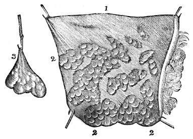
Fig. 4. 1, A portion of the adipose tissue. 2, 2, 2, Minute bags containing fat. 3, A cluster of these bags, separated and suspended.
37. The CARTILAGINOUS TISSUE is firm, smooth, and highly elastic. Except bone, it is the hardest part of the animal frame. It tips the ends of the bones that concur in forming a joint. Its use is to facilitate the motion of the joints by its smooth surface, while its elastic character diminishes the shock that would otherwise be experienced if this tissue were inelastic.
36. Describe the adipose tissue. Where does this tissue principally exist? Give observation in regard to the adipose tissue. 37. Describe the cartilaginous tissue. What is its use?
38 The OSSEOUS TISSUE, in composition and arrangement of matter, varies at different periods of life, and in different bones. In some instances, the bony matter is disposed in plates, while in other instances, the arrangement is cylindrical. Sometimes, the bony matter is dense and compact; again, it is spongy, or porous. In the centre of the long bones, a space is left which is filled with a fatty substance, called mar´row.
Observation. Various opinions exist among physiologists in regard to the use of marrow. Some suppose it serves as a reservoir of nourishment, while others, that it keeps the bones from becoming dry and brittle. The latter opinion, however, has been called in question, as the bones of the aged man contain more marrow than those of the child, and they are likewise more brittle.
Fig. 5.
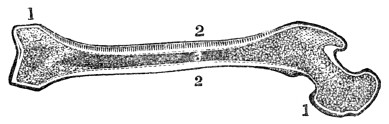
Fig.
5. A section of the femur, (thigh-bone.) 1, 1, The extremities, showing a thin plate of compact texture, which covers small cells, that diminish in size, but increase in number, as they approach the articulation. 2, 2, The walls of the shaft, which are very firm and solid. 3, The cavity that contains the marrow.39. The MUSCULAR TISSUE is composed of many fibres, that unite to form fasciculi, each of which is enclosed in a delicate layer of cellular tissue. Bundles of these fasciculi constitute a muscle.
Observation. A piece of boiled beef will clearly illustrate the arrangement of muscular fibre.
38. What is said of the osseous tissue? How is the bony matter arranged in different parts of the animal frame? What is said of the use of marrow? 39. Of what is the muscular tissue composed? How may the arrangement of muscular fibre be illustrated?
40. The MUCOUS TISSUE differs from the serous by its lining all the cavities which communicate with the air. The nostrils, the mouth, and the stomach afford examples. The external surface of this membrane, or that which is exposed to the air, is soft, and bears some resemblance to the downy rind of a peach. It is covered by a viscid fluid called mu´cus. This is secreted by small gland-cells, called ep-i-the´li-a, or secretory cells of the mucous membrane. The use of this membrane and its secreted mucus is to protect the inner surface of the cavities which it lines.
Observation. A remarkable sympathy exists between the remote parts of the mucous membrane. Thus the condition of the stomach may be ascertained by an examination of the tongue.
41. The NERVOUS TISSUE consists of soft, pulpy matter, enclosed in a sheath, called neu-ri-lem´a. This tissue consists of two substances. The one, of a pulpy character and gray color, is called cin-e-ri´tious, (ash-colored.) The other, of a fibrous character and white, is named med´ul-la-ry, (marrow-like.) In every part of the nervous system both substances are united, with the exception of the nervous fibres and filaments, which are solely composed of the medullary matter enclosed in a delicate sheath.
40. How does the mucous differ from the
serous
tissue? What is the appearance of the external surface of this membrane? Where is the mucus secreted? What is the use of this membrane? 41. Of what does the nervous tissue consist? Describe the two substances that enter into the composition of the nervous tissue.CHAPTER III
CHEMISTRY OF THE HUMAN BODY.
42. An ULTIMATE ELEMENT is the simplest form of matter with which we are acquainted; as gold, iron, &c.
43. These elements are divided into metallic and non-metallic substances. The metallic substances are Po-tas´si-um, So´di-um, Cal´ci-um, Mag-ne´si-um, A-lu´min-um, I´ron, Man´ga-nese, and Cop´per. The non-metallic substances are Ox´y-gen, Hy´dro-gen, Car´bon, Ni´tro-gen, Si-li´-ci-um, Phos´phor-us, Sul´phur, Chlo´rine, and a few others.
44. Potash (potassium united with oxygen) is found in the blood, bile, perspiration, milk, &c.
45. Soda (sodium combined with oxygen) exists in the muscles, and in the same fluids in which potash is found.
46. Lime (calcium combined with oxygen) forms the principal ingredient of the bones. The lime in them is combined with phosphoric and carbonic acid.
47. Magnesia (magnesium combined with oxygen) exists in the bones, brain, and in some of the animal fluids; as milk.
48. Silex (silicium combined with oxygen) is contained in the hair and in some of the secretions.
49. Iron forms the coloring principle of the red globules of the blood, and is found in every part of the system.
Observation. As metallic or mineral substances enter into the ultimate elements of the body, the assertion that all minerals are poisonous, however small the quantity, is untrue.
42. What is an ultimate element? Give examples. 43. How are they divided? Name the metallic substances. Name the non-metallic substances. 44. What is said of potash? 45. Of soda? 46. Of lime? 47. Of magnesia? 48. Of silex? 49. What forms the coloring principle of the blood? What is said of mineral substances?
50. Oxygen is contained in all the fluids and solids of the body. It is almost entirely derived from the inspired air and water. It is expelled in the form of carbonic acid and water from the lungs and skin. It is likewise removed in the other secretions.
51. Hydrogen is found in all the fluids and in all the solids of the body. It is derived from the food, as well as from water and other drinks. It exists in the greatest abundance in the impure, dark-colored blood of the system. It is removed by the agency of the kidneys, skin, lungs, and other excretory organs.
52. Carbon is an element in the oil, fat, albumen, fibrin, gelatin, bile, and mucus. This element likewise exists in the impure blood in the form of carbonic acid gas. Carbon is obtained from the food, and discharged from the system by the secretions and respiration.
53. Nitrogen is contained in most animal matter, but is most abundant in fibrin. It is not contained in fat and a few other substances.
Observation. The peculiar smell of animal matter when burning is owing to nitrogen. This element combined with hydrogen forms am-mo´ni-a, (hartshorn,) when animal matter is in a state of putrefaction.
54. Phosphorus is contained in many parts of the system, but more particularly in the bones. It is generally found in combination with oxygen, forming phosphoric acid. The phosphoric acid is usually combined with alkaline bases; as lime in the bones, forming phosphate of lime.
55. Sulphur exists in the bones, muscles, hair, and nails. It is expelled from the system by the skin and intestines.
56. Chlorine is found in the blood, gastric juice, milk, perspiration, and saliva.
50. What is said of oxygen? 51. Of hydrogen? 52. What is said of carbon? 53. Of nitrogen? How is ammonia formed? 54. What is said of phosphorus? 55. What is said of sulphur? 56. Of chlorine?
57. Proximate elements are forms of matter that exist in organized bodies in abundance, and are composed chiefly of oxygen, hydrogen, carbon, and nitrogen, arranged in different proportions. They exist already formed, and may be separated in many instances, by heat or mechanical means. The most important compounds are Al-bu´men,
Fi´brin,
Gel´a-tin, Mu´cus, Fat, Ca´se-ine, Chon´drine, Lac´tic acid, and Os´ma-zome.58. Albumen is found in the body, both in a fluid and solid form. It is an element of the skin, glands, hair, and nails, and forms the principal ingredient of the brain. Albumen is without color, taste, or smell, and it coagulates by heat, acids, and alcohol.
Observation. The white of an egg is composed of albumen, which can be coagulated or hardened by alcohol. As albumen enters so largely into the composition of the brain, is not the impaired intellect and moral degradation of the inebriate attributable to the effect of alcohol in hardening the albumen of this organ?
59. Fibrin exists abundantly in the blood, chyle, and lymph. It constitutes the basis of the muscles. Fibrin is of a whitish color, inodorous, and insoluble in cold water. It differs from albumen by possessing the property of coagulating at all temperatures.
Observation. Fibrin may be obtained by washing the thick part of blood with cold water; by this process, the red globules, or coloring matter, are separated from this element.
60. Gelatin is found in nearly all the solids, but it is not known to exist in any of the fluids. It forms the basis of the cellular tissue, and exists largely in the skin, bones, ligaments, and cartilages.
57. What are proximate elements? Do they exist already formed in
organized
bodies? Name the most important compounds. 58. What is said of albumen? Give observation relative to this element. 59. Of fibrin? How does albumen differ from fibrin? How can fibrin be obtained? 60. What is said of gelatin?Observation. Gelatin is known from other organic principles by its dissolving in warm water, and forming “jelly.” When dry, it forms the hard, brittle substance, called glue. Isinglass, which is used in the various mechanical arts, is obtained from the sounds of the sturgeon.
61. Mucus is a viscid fluid secreted by the gland-cells, or epithelia. Various substances are included under the name of mucus. It is generally alkaline, but its true chemical character is imperfectly understood. It serves to moisten and defend the mucous membrane. It is found in the cuticle, brain, and nails; and is scarcely soluble in water, especially when dry. (Appendix C.)
62. Osmazome is a substance of an aromatic flavor. It is of a yellowish-brown color, and is soluble both in water and alcohol, but does not form a jelly by concentration. It is found in all the fluids, and in some of the solids; as the brain.
Observation. The characteristic odor and taste of soup are owing to osmazome.
63. There are several acids found in the human system; as the A-ce´tic, Ben-zo´ic, Ox-al´ic, U´ric, and some other substances, but not of sufficient importance to require a particular description.
How is it known from other organic principles? 61. What is said of mucus? 62. Of osmazome? To what are the taste and odor of soup owing? 63. What acids are found in the system?
CHAPTER IV.
THE BONES.
64. The bones are firm and hard, and of a dull white color. In all the higher orders of animals, among which is man, they are in the interior of the body, while in lobsters, crabs, &c., they are on the outside, forming a case which protects the more delicate parts from injury.
65. In the mechanism of man, the variety of movements he is called to perform requires a correspondent variety of component parts, and the different bones of the system are so admirably adapted to each other, that they admit of numerous and varied motions.
66. When the bones composing the skeleton are united by natural ligaments, they form what is called a natural skeleton, when united by wires, what is termed an artificial skeleton.
67. The elevations, or protuberances, of the bones are called proc´es-ses, and are, generally, the points of attachment for the muscles and ligaments.
ANATOMY OF THE BONES.
68. The BONES are composed of both animal and earthy matter. The earthy portion of the bones gives them solidity and strength, while the animal part endows them with
vitality.
64. What is said of the bones? 65. Is there an adaptation of the bones of the system to the offices they are required to perform? 66. What is a natural skeleton? What an artificial? 67. What part of the bones are called processes? 68–73. Give the structure of the bones. 68. Of what are the bones composed? What are the different uses of the component parts of the bones?
Experiments. 1st. To show the earthy without the animal matter, burn a bone in a clear fire for about fifteen minutes, and it becomes white and brittle, because the gelatin, or animal matter of the bone, has been destroyed.
2d. To show the animal without the earthy matter of the bones, immerse a slender bone for a few days in a weak acid, (one part muriatic acid and six parts water,) and it can then be bent in any direction. In this experiment, the acid has removed the earthy matter, (carbonate and phosphate of lime,) yet the form of the bone is unchanged.
69. The bones are formed from the blood, and are subjected to several changes before they are perfected. At their early formative stage, they are cartilaginous. The vessels of the cartilage, at this period, convey only the lymph, or white portion of the blood; subsequently, they convey red blood. At this time, true ossification (the deposition of phosphate and carbonate of lime) commences at certain points, which are called the points of ossification.
70. Most of the bones are formed of several pieces, or centres of ossification. This is seen in the long bones which have their extremities separated from the body by a thin partition of cartilage. It is some time before these separate pieces are united to form one bone.
71. When the process of ossification is completed, there is still a constant change in the bones. They increase in bulk, and become less vascular, until middle age. In advanced life, the elevations upon their surface and near the extremities become more prominent, particularly in individuals accustomed to labor. As a person advances in years, the vitality diminishes, and in extreme old age, the earthy substance predominates; consequently, the bones are extremely brittle.
How can the earthy matter of the bones be shown? The animal? 69. What is the appearance of the bones in their early formative stage? When does true ossification commence? 70. How are most of the bones formed? 71. What is said of the various changes of the bones after ossification?
72. The fibrous membrane that invests the bones is called per-i-os´te-um; that which covers the cartilages is called per-i-chon´dri-um. When this membrane invests the skull, it is called per-i-cra´ni-um.
Fig. 6.
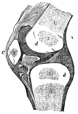
Fig. 6. A section of the knee-joint. The lower part of the femur, (thigh-bone,) and upper part of the tibia, (leg-bone,) are seen ossified at 1, 1. The cartilaginous extremities of the two bones are seen at d, d. The points of ossification of the extremities, are seen at 2, 2. The patella, or knee-pan, is seen at c. 3, A point, or centre of ossification.
73. The PERIOSTEUM is a firm membrane immediately investing the bones, except where they are tipped with cartilage, and the crowns of the teeth, which are protected by enamel. This membrane has minute nerves, and when healthy, possesses but little sensibility. It is the nutrient membrane of the bone, endowing its exterior with vitality; it also gives insertion to the tendons and connecting ligaments of the joints.
72. What is the membrane called that invests the bones? That covers the cartilage? That invests the skull? Explain fig. 6. 73. Describe the periosteum.
74. There are two hundred and eight[3] bones in the human body, beside the teeth. These, for convenience, are divided into four parts: 1st. The bones of the Head. 2d. The bones of the Trunk. 3d. The bones of the Upper Extremities. 4th. The bones of the Lower Extremities.
75. The bones of the HEAD are divided into those of the Skull, Ear, and Face.
76. The SKULL is composed of eight bones. They are formed of two plates, or tablets of bony matter, united by a porous portion of bone. The external tablet is fibrous and tough; the internal plate is dense and hard, and is called the vit´re-ous, or glassy table. These tough, hard plates are adapted to resist the penetration of sharp instruments, while the different degrees of density possessed by the two tablets, and the intervening spongy bone, serve to diminish the vibrations that would occur in falls or blows.
77. The skull is convex externally, and at the base much thicker than at the top or sides. The most important part of the brain is placed here, completely out of the way of injury, unless of a very serious nature. The base of the cranium, or skull, has many projections, depressions, and apertures; the latter affording passages for the nerves and blood-vessels.
74. How many bones in the human body? How are they divided? 75–81. Give the anatomy of the bones of the head. 75. How are the bones of the head divided? 76. Describe the bones of the skull. 77. What is the form of the skull? What does the base of the skull present?
78. The bones of the cranium are united by ragged edges, called sut´ures. The edges of each bone interlock with each other, producing a union, styled, in carpentry, dovetailing. They interrupt, in a measure, the vibrations produced by external blows, and also prevent fractures from extending as far as they otherwise would, in one continued bone. From infancy to the twelfth year, the sutures are imperfect; but, from that time to thirty-five or forty, they are distinctly marked; in old age, they are nearly obliterated.
Fig. 7.
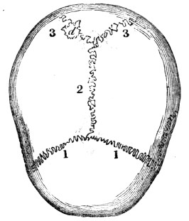
Fig.
7.
1, 1, The coronal suture at the front and upper part of the skull, orcranium
. 2, The sagittal suture on the top of the skull. 3, 3, The lambdoidal suture at the back part of the cranium.79. We find as great a diversity in the form and texture of the skull-bone, as in the expression of the face. The head of the New Hollander is small; that of the African is compressed; while the Caucasian is distinguished for the beautiful oval form of the head. The Greek skulls, in texture, are close and fine, while the Swiss are softer and more open.
78. How are the bones of the skull united? What are the uses of the sutures? Mention the appearance of the sutures at different ages. What does fig. 7 represent? 79. What is said respecting the form and texture of the skull in different nations?
80. In each EAR are four very small bones. They aid in hearing.
81. In the FACE are fourteen bones, some of which serve for the attachment of powerful muscles, which are more or less called into action in masticating food; others retain in place the soft parts of the face.
Fig. 8.
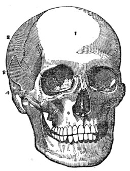
Fig. 8. 1, The frontal, or bone of the forehead. 2. The parietal bone. 3, The temporal bone. 4, The zygomatic process of the temporal bone. 5, The malar (cheek) bone. 6, The superior maxillary bone, (upper jaw.) 7, The vomer, that separates the cavities of the nose. 8, The inferior maxillary bone, (lower jaw.) 9. The cavity for the eye.
82. The TRUNK has fifty-four bones—twenty-four Ribs; twenty-four bones in the Spi´nal Col´umn, (back-bone;) four in the Pel´vis; the Ster´num, (breast-bone;) and the Os hy-oid´es, (the bone at the base of the tongue.) They are so arranged as to form, with the soft parts attached to them, two cavities, called the Tho´rax (chest) and Ab-do´men.
80. How many bones in the ear? 81. How many bones in the face? What is their use? Explain fig. 8. 82–94. Give the anatomy of the bones of the trunk. 82. How many bones in the trunk? Name them. What do they form by their arrangement?
83. The THORAX is formed by the sternum in front; the ribs, at the sides; and the twelve dorsal bones of the spinal column, posteriorly. The natural form of the chest is a cone, with its apex above; but fashion, in many instances, has nearly inverted this order. This cavity contains the lungs, heart, and large blood-vessels.
Fig. 9.
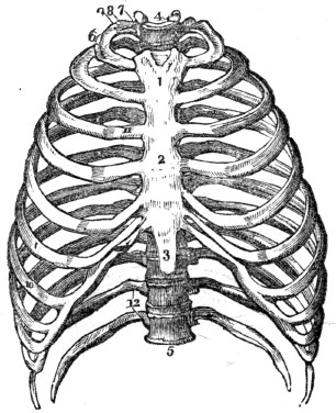
Fig.
9. 1, The first bone of the sternum, (breast-bone.) 2. The second bone of the sternum. 3, The cartilage of the sternum. 4, The first dorsal vertebra, (a bone of the spinal column.) 5, The last dorsal vertebra. 6, The first rib. 7, Its head. 8, Its neck. 9, Its tubercle. 10, The seventh, or last true rib. 11, The cartilage of the third rib. 12, The floating ribs.84. The STERNUM is composed of eight pieces in the child. These unite and form but three parts in the adult. In youth, the two upper portions are converted into bone, while the lower portion remains cartilaginous and flexible until extreme old age, when it is often converted into bone.
85. The RIBS are connected with the spinal column, and increase in length as far as the seventh. From this they successively become shorter. The direction of the ribs from above, downward, is oblique, and their curve diminishes from the first to the twelfth. The external surface of each rib is convex; the internal, concave. The inferior, or lower ribs, are, however, very flat.
83. Describe the thorax. Explain fig. 9. 84. Describe the
sternum.
85. Describe the ribs.86. The seven upper ribs are united to the sternum, through the medium of cartilages, and are called the true ribs. The cartilages of the next three are united with each other, and are not attached to the sternum; these are called false ribs. The lowest two are called floating ribs, as they are not connected either with the sternum or the other ribs.
87. The SPINAL COLUMN is composed of twenty-four pieces of bone. Each piece is called a vert´e-bra. On examining one of the bones, we find seven projections, called processes; four of these, that are employed in binding the bones together, are called articulating processes; two of the remaining are called the transverse; and the other, the spinous. The last three give attachment to the muscles of the back.
88. The large part of the vertebra, called the body, is round and spongy in its texture, like the extremity of the round bones. The processes are of a more dense character. The projections are so arranged that a tube, or canal, is formed immediately behind the bodies of the vertebræ, in which is placed the me-dul´la spi-na´lis, (spinal cord,) sometimes called the pith of the back-bone.
89. Between these joints, or vertebræ, is a peculiar and highly elastic substance, which much facilitates the bending movements of the back. This compressible cushion of cartilage also serves the important purpose of diffusing and diminishing the shock in walking, running, or leaping, and tends to protect the delicate texture of the brain.
86. How are the ribs united to the sternum? 87. Describe the spinal column. 88. Give the structure of the vertebra. Where is the spinal cord placed? 89. What is placed between each
vertebra?
What is its use?90. Another provision for the protection of the brain, which bears convincing proof of the wisdom and beneficence of the Creator, is the antero-posterior, or forward and backward curve of the spinal column. Were it a straight column, standing perpendicularly, the slightest jar, in walking, would cause it to recoil with a sudden jerk; because, the weight bearing equally, the spine would neither yield to the one side nor the other. But, shaped as it is, we find it yielding in the direction of the curves, and thus the force of the shock is diffused.
Fig. 10.
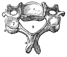
Fig. 10. A vertebra of the neck. 1, The body of the vertebra. 2, The spinal canal. 4, The spinous process, cleft at its extremity. 5, The transverse
process.
7, The inferior articulating process. 8, The superior articulating process.Fig. 11.
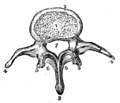
Fig. 11. 1, The cartilaginous substance that connects the bodies of the vertebræ. 2, The body of the vertebra. 3, The spinous process. 4, 4, The transverse processes. 5, 5, The articulating processes. 6, 6, A portion of the bony bridge that assists in forming the spinal canal, (7.)
Observation. A good idea of the structure of the vertebræ may be obtained by examining the spinal column of a domestic animal, as the dog, cat, or pig.
91. The PELVIS is composed of four bones; the two in-nom-i-na´ta, (nameless bones,) the sa´crum, and the coc´cyx.
92. The INNOMINATUM, in the child, consists of three pieces. These, in the adult, become united, and constitute but one bone. In the sides of these bones is a deep socket, or depression, like a cup, called the ac-e-tab´u-lum, in which the round head of the thigh-bone is placed.
90. What is said of the curves of the spinal column? What is represented by fig. 10? By fig. 11? How can the structure of the vertebræ be seen? 91. Of how many bones is the pelvis composed? 92. What is said of the innominatum in the child?
93. The SACRUM, so called because the ancients offered it in sacrifices, is a wedge-shaped bone, that is placed between the innominata, and to which it is bound by ligaments. Upon its upper surface it connects with the lower vertebra. At its inferior, or lower angle, it is united to the coccyx. It is concave upon its anterior, and convex upon its posterior surface.
Fig. 12.
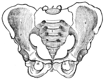
Fig. 12. 1, 1, The innominata, (nameless bones.) 2, The sacrum. 3, The
coccyx.
4, 4, The acetabulum. a, a, The pubic portion of the innominata. d, The arch of the pubes; e, The junction of the sacrum and lower lumbar vertebra.94. The COCCYX, in infants, consists of several pieces, which, in youth, become united and form one bone. This is the terminal extremity of the spinal column.
In the adult? Describe the acetabulum. 93. Describe the
sacrum.
Explain fig. 12. 94. Describe the coccyx.CHAPTER V.
ANATOMY OF THE BONES, CONTINUED
95. The bones of the upper and lower limbs are enlarged at each extremity, and have projections, or processes. To these, the tendons of muscles and ligaments are attached, which connect one bone with another. The shaft of these bones is cylindrical and hollow, and in structure, their exterior surface is hard and compact, while the interior portion is of a reticulated character. The enlarged extremities of the round bones are more porous than the main shaft.
96. The UPPER EXTREMITIES contain sixty-four bones—the Scap´u-la, (shoulder-blade;) the Clav´i-cle, (collar-bone;) the Hu´mer-us, (first bone of the arm;) the Ul´na and Ra´di-us, (bones of the fore-arm;) the Car´pus, (wrist;) the Met-a-car´pus, (palm of the hand;) and the Pha-lan´ges, (fingers and thumb.)
97. The CLAVICLE is attached, at one extremity, to the sternum; at the other, it is united to the scapula. It is shaped like the Italic ∫. Its use is to keep the arms from sliding toward the breast.
98. The SCAPULA is situated upon the upper and back part of the chest. It is flat, thin, and of a triangular form. This bone lies upon and is retained in its position by muscles. By their contractions it may be moved in different directions.
99. The HUMERUS is cylindrical, and is joined at the elbow with the ulna of the fore-arm; at the scapular extremity, it is lodged in the glenoid cavity, where it is surrounded by a membranous bag, called the capsular ligament.
95–104. Give the anatomy of the bones of the upper extremities. 95. Give the structure of the bones of the extremities. 96. How many bones in the upper extremities? Name them. 97. Give the attachments of the clavicle. What is its use? 98. Describe the scapula. How is it retained in its position? 99. Describe the humerus.
Fig. 13.
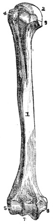
Fig. 13. 1, The shaft of the humerus. 2, The large, round head that is placed in the glenoid cavity. 3, 4, Processes, to which muscles are attached. 5, A process, called the external elbow. 6, A process, called the internal elbow. 7, The articulating surface upon which the ulna rolls.
Fig. 14.
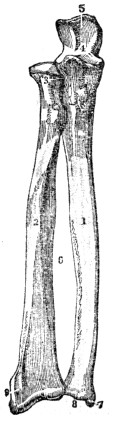
Fig. 14. 1, The body of the ulna. 2, The shaft of the radius. 3, The upper articulation of the radius and ulna. 4, Articulating cavity, in which the lower extremity of the humerus is placed. 5, Upper extremity of the ulna, called the olecranon process, which forms the point of the elbow. 6, Space between the radius and ulna, filled by the intervening ligament. 7, Styloid process of the ulna. 8, Surface of the radius and the ulna, where they articulate with the bones of the wrist. 9, Styloid process of the radius.
100. The ULNA articulates with the humerus at the elbow, and forms a perfect hinge-joint. This bone is situated on the inner side of the fore-arm.
What is represented by fig. 13? By fig. 14? 100. Describe the ulna.
101. The RADIUS articulates with the bones of the carpus and forms the wrist-joint. This bone is situated on the
outside
of the fore-arm, (the side on which the thumb is placed.) The ulna and radius, at their extremities, articulate with each other, by which union the hand is made to rotate, permitting its complicated and varied movements.102. The CARPUS is composed of eight bones, ranged in two rows, and so firmly bound together, as to permit only a small amount of movement.
Fig. 15.
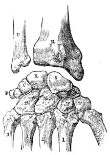
Fig. 15. U, The ulna. R, The radius. S, The scaphoid bone. L, The semilunar bone. C, The cuneiform bone. P, The pisiform bone. These four form the first row of carpal bones. T, T, The trapezium and trapezoid bones. M, The os magnum. U, The unciform bone. These four form the second row of carpal bones. 1, 1, 1, 1, 1, The metacarpal bones of the thumb and fingers.
Fig. 16.
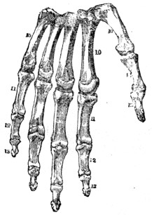
Fig. 16. 10, 10, 10, The metacarpal bones of the hand. 11, 11, First range of finger-bones. 12, 12, Second range of finger-bones. 13, 13, Third range of finger-
bones.
14, 15, Bones of the thumb.103. The METACARPUS is composed of five bones, upon four of which the first range of the finger-bones is placed; and upon the other, the first bone of the thumb. The five
metacarpal
bones articulate with the second range of carpal bones.101. The radius. 102. How many bones in the carpus? How are they ranged?
103.
Describe themetacarpus.
104. The PHALANGES of the fingers have three ranges of bones, while the thumb has but two.
Observation. The wonderful adaptation of the hand to all the mechanical offices of life, is one cause of man’s superiority over the rest of creation. This arises from the size and strength of the thumbs, and the different lengths of the fingers.
105. The LOWER EXTREMITIES contain sixty bones—the Fe´mur, (thigh-bone;) the Pa-tel´la, (knee-pan;) the Tib´i-a, (shin-bone;) the Fib´u-la, (small bone of the leg;) the Tar´sus, (instep;) the Met-a-tar´sus, (middle of the foot;) and the Pha-lan´ges, (toes.)
106. The FEMUR is the longest bone in the system. It supports the weight of the head, trunk, and upper extremities. The large, round head of this bone is placed in the acetabulum. This articulation is a perfect specimen of the ball and socket joint.
107. The PATELLA is a small bone connected with the tibia by a strong ligament. The tendon of the ex-tens´or muscles of the leg is attached to its upper edge. This bone is placed on the anterior part of the lower extremity of the femur, and acts like a pulley, in the extension of the limb.
108. The TIBIA is the largest bone of the leg. It is of a triangular shape, and enlarged at each extremity.
109. The FIBULA is a smaller bone than the tibia, but of
similar
shape. It is firmly bound to the tibia, at each extremity.110. The TARSUS is formed of seven irregular bones, which are so firmly bound together as to permit but little movement.
104. How many ranges of bones have the phalanges? 105–112. Give the anatomy of the bones of the lower extremities. 105. How many bones in the lower extremities? Name them. 106. Describe the femur. 107. Describe the patella. What is its function? 108. What is the largest bone of the leg called? What is its form? 109. What is said of the fibula? 110. Describe the tarsus.
Fig. 17.
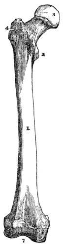
Fig.
17. 1, The shaft of the femur, (thigh-bone.) 2, A projection, called the trochantar minor, to which are attached some strong muscles. 4, The trochantar major, to which the large muscles of the hip are attached. 3, The head of the femur. 5, The external projection of the femur, called the external condyle. 6, The internal projection, called the internal condyle. 7, The surface of the lower extremity of the femur, that articulates with the tibia, and upon which the patella slides.Fig. 18.
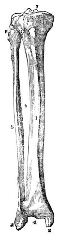
Fig. 18. 1, The tibia. 5, The fibula. 8, The space between the two, filled with the inter-osseous ligament. 6, The junction of the tibia and fibula at their upper extremity. 2, The external malleolar process, called the external ankle. 3, The internal malleolar process, called the internal ankle. 4, The surface of the lower extremity of the tibia, that unites with one of the tarsal bones to form the ankle-joint. 7, The upper extremity of the tibia, upon which the lower extremity of the femur rests.
Explain fig. 17. Explain fig. 18.
111. The METATARSAL bones are five in number. They articulate at one extremity with one range of tarsal bones;
at
the other extremity, with the first range of the toe-bones.Fig. 19.
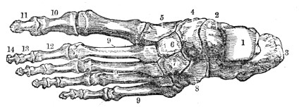
Fig. 19. A representation of the upper surface of the bones of the foot. 1, The surface of the astragulus, where it unites with the tibia. 2, The body of the astragulus. 3, The calcis, (heel-bone.) 4, The scaphoid bone. 5, 6, 7, The cuneiform bones. 8, The cuboid. 9, 9, 9, The metatarsal bones. 10, The first bone of the great toe. 11, The second bone. 12, 13, 14, Three ranges of bones, forming the small toes
Fig. 20.
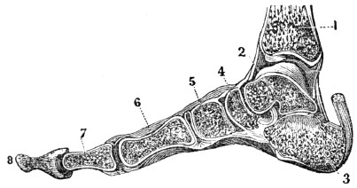
Fig. 20. A side view of the bones of the foot, showing its arched form. The arch rests upon the heel behind, and the ball of the toes in front. 1, The lower part of the tibia. 2, 3, 4, 5, Bones of the tarsus. 6, The metatarsal bone. 7, 8, The bones of the great toe. These bones are so united as to secure a great degree of elasticity, or spring.
Observation. The tarsal and metatarsal bones are united so as to give the foot an arched form, convex above, and concave below. This structure conduces to the elasticity of the step, and the weight of the body is transmitted to the ground by the spring of the arch, in a manner which prevents injury to the numerous organs.
111. Describe the metatarsal bones. Explain fig. 19. What is represented by fig. 20? What is said of the arrangement of the bones of the foot?
112. The PHALANGES (fig. 19) are composed of fourteen bones; each of the small toes has three ranges of bones, while the great toe has but two.
113. The JOINTS form an interesting part of the body. In their construction, every thing shows the regard that has been paid to the security and the facility of motion of the parts thus connected together. They are composed of the extremities of two or more bones, Car´ti-lages, (gristles,) Syn-o´vi-al membrane, and Lig´a-ments.
[3]
Some anatomists reckon more than this number, others less, for the reason that, at different periods of life, the number of pieces of which one bone is formed, varies. Example. The breast-bone, in infancy, has eight pieces; in youth, three; in old age, but one.
Fig. 21.
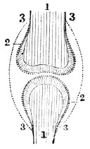
Fig. 21 The relative position of the bones, cartilages, and synovial
membrane.
1, 1, The extremities of two bones that concur to form a joint. 2, 2, The cartilages that cover the end of the bones. 3, 3, 3, 3, The synovial membrane which covers the cartilage of both bones, and is then doubled back from one to the other; it is represented by the dotted lines.Fig. 22.
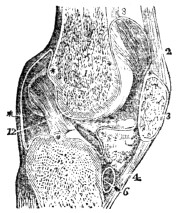
Fig. 22. A vertical section of the knee-joint. 1, The femur. 3, The patella. 5, The tibia. 2, 4, The ligaments of the patella. 6, The cartilage of the tibia 12, The cartilage of the femur. * * * *, The synovial membrane.
114. Cartilage is a smooth, solid, elastic substance, of a pearly whiteness, softer than bone. It forms upon the articular surfaces of the bones a thin incrustation, not more than the sixteenth of an inch in thickness. Upon convex surfaces it is the thickest in the centre, and thin toward the circumference; while upon concave surfaces, an opposite arrangement is presented.
112. Describe the phalanges. 113–118. Give the anatomy of the
joints.
113. What is said of the joints? Of what are the jointscomposed?
What is illustrated by fig. 21? By fig. 22? 114. Define cartilage.115. The SYNOVIAL MEMBRANE is a thin, membranous layer, which covers the cartilages, and is thence bent back, or reflected upon the inner surfaces of the ligaments which surround and enter into the composition of the joints. This membrane forms a closed sac, like the membrane that lines an egg-shell.
Fig. 23.
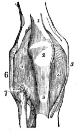
Fig. 23. The anterior ligaments of the knee-joint. 1, The tendon of the muscle that extends the leg. 2, The patella. 3, The anterior ligament of the patella, near its insertion. 4, 4, The synovial membrane. 5, The internal lateral ligament. 6, The long external lateral ligament. 7, The anterior and superior ligament that unites the fibula to the tibia.
Fig. 24.
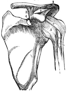
Fig. 24. 2, 3, The ligaments that extend from the clavicle (1) to the scapula (4.) The ligaments 5, 6, extend from the scapula to the first bone of the arm.
116. Beside the synovial membrane, there are numerous smaller sacs, called bur´sæ mu-co´sæ. These are often associated with the articulation. In structure, they are analogous to synovial membranes, and secrete a similar fluid.
115. Describe the synovial membrane. 116. Describe the bursæ mucosæ. What is represented by fig. 23? By fig. 24?
117. The LIGAMENTS are composed of numerous straight fibres, collected together, and arranged into short bands of various breadths, or so interwoven as to form a broad layer, which completely surrounds the articular extremities of the bones, and constitutes a capsular ligament. These connecting bands are white, glistening, and inelastic. Most of the ligaments are found exterior to the synovial membrane.
118. The bones, cartilages, ligaments, and synovial membrane are insensible when in health; yet they are supplied with organic nerves, as well as with arteries, veins, and lymphatics.
Observation. The joints of the domestic animals are similar in their construction to those of man. To illustrate this part of the body, a fresh joint of the calf or sheep may be used. After divesting the joints of the skin, the satin-like bands, or ligaments, will be seen passing from one bone to the other, under which may be observed the membranous bag, called the capsular ligament. This is very smooth, as it is lined with the soft synovial membrane, beneath which will be seen the cartilage, that may be cut with a knife, and under this the rough extremity of the ends of the bones.
117. Of what are ligaments composed? What is the appearance of these bands? Where are they found? 118. With what vessels are the cartilages and ligaments supplied? How can the structure of the joints be explained?
CHAPTER VI.
PHYSIOLOGY OF THE BONES.
119. The bones are the framework of the system. By their solidity and form, they not only retain every part of the fabric in its proper shape, but afford a firm surface for the attachment of the muscles and ligaments. By means of the bones, the human frame presents to the eye a wonderful piece of mechanism, uniting the most finished symmetry of form with freedom of motion, and also giving security to many important organs.
120. To give a clear idea of the relative uses of the bones and muscles, we will quote the comparison of another, though, as in other comparisons, there are points of difference. The “bones are to the body what the masts and spars are to the ship,—they give support and the power of resistance. The muscles are to the bones what the ropes are to the masts and spars. The bones are the levers of the system; by the action of the muscles their relative positions are changed. As the masts and spars of a vessel must be sufficiently firm to sustain the action of the ropes, so the bones must possess the same quality to sustain the action of the muscles in the human body.”
121. Some of the bones are designed exclusively for the protection of the organs which they enclose. Of this number are those that form the skull, the sockets of the eye, and the cavity of the nose. Others, in addition to the protection they give to important organs, are useful in movements of certain kinds. Of this class are the bones of the spinal column, and ribs. Others are subservient to motion. Of this class are the upper and lower extremities.
119–128. Give the physiology of the bones. 119. How may the bones be considered? 120. To what may the bones be compared? 121. Give the different offices of the bones.
122. The bones are subject to growth and decay; to removal of old, useless matter, and the deposit of new particles, as in other tissues. This has been tested by the following experiment. Some of the inferior animals were fed with food that contained madder. In a few days, some of the animals were killed, and their bones exhibited an unusually reddish appearance. The remainder of the animals were, for a few weeks, fed on food that contained no coloring principle. When they were killed, their bones exhibited the usual color of such animals. The coloring matter, which had been deposited, had been removed by the action of the lymphatics.
123. The extremities of the bones that concur in forming a joint, correspond by having their respective configurations reciprocal. They are, in general, the one convex, and the other concave. In texture they are porous, and consequently more elastic than if more compact. These are covered with a cushion of cartilage. The elastic character of these parts acts as so many springs, in diminishing the jar which important organs of the system would otherwise receive.
124. The synovial membrane secretes a viscous fluid, which is called syn-o´vi-a. This lubricating fluid of the joints enables the surfaces of the bones and tendons to move smoothly upon each other, thus diminishing the friction consequent on their action.
Observations. 1st. In this secretion is manifested the skill and omnipotence of the Great Architect; for no machine of human invention supplies to itself, by its own operations, the necessary lubricating fluid. But, in the animal frame, it is supplied in proper quantities, and applied in the proper place, and at the proper time.
122. What is said of the change in bones? How was it proved that there was a constant change in the osseous fabric? 123. What is said of the extremities of the bones that form a joint? 124. What is synovia? Its use? What is said of this lubricating fluid?
2d. In some cases of injury and disease, the synovial fluid is secreted in large quantities, and distends the sac of the joint. This affection is called dropsy of the joint, and occurs most frequently in that of the knee.
125. The function of the ligaments is to connect and bind together the bones of the system. By them the small bones of the wrist and foot, as well as the large bones, are as securely fastened as if retained by clasps of steel. Some of them are situated within the joints, like a central cord, or pivot, (3, fig. 26.) Some surround it like a hood, and contain the lubricating synovial fluid, (8, 9, fig. 25,) and some in the form of bands at the side, (5, 6, fig. 23.)
Fig. 25.
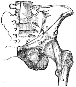
Fig. 25. 8, 9, The ligaments that extend from the hip-bone (6) to the femur, (5.)
Fig. 26.
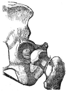
Fig. 26. 2, The socket of the hip-joint. 5, The head of the femur, which is lodged in the socket. 3, The ligament within the socket.
126. By the ligaments the lower jaw is bound to the temporal bones, and the head to the neck. They extend the whole length of the spinal column, in powerful bands, on the outer surface, between the spinal bones, and from one spinous process to another. They bind the ribs to the vertebræ, to the transverse process behind, and to the sternum in front; and this to the clavicle; and this to the first rib and scapula; and this last to the humerus.
What is the effect when the synovial fluid is secreted in large quantities? 125. What is the function of the ligaments? 126. Mention how the bones of the system are connected.
127. They also bind the two bones of the fore-arm at the elbow-joint; and these to the wrist; and these to each other and to those of the hand; and these last to each other and to those of the fingers and thumb. In the same manner, they bind the bones of the pelvis together; and these to the femur; and this to the two bones of the leg and patella; and so on, to the ankle, foot, and toes, as in the upper extremities.
Fig. 27.
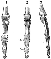
Fig. 27. 1, A front view of the lateral ligaments of the finger-joints. 2, A view of the anterior ligaments (a, b,) of the finger-joints. 3, A side view of the lateral ligaments of the finger joints.
128. The different joints vary in range of movement, and in complexity of structure. Some permit motions in all directions, as the shoulder; some move in two directions, permitting only flexion and extension of the part, as the elbow; while others have no movement, as the bones of the head in the adult.
Explain fig. 27. 128. Describe the variety of movements in the different joints.
Fig. 28
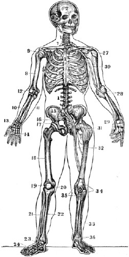
Fig. 28. 1, 1, The spinal column. 2, The skull. 3, The lower jaw. 4, The sternum. 5, The ribs. 6, 6, The cartilages of the ribs. 7, The clavicle. 8, The humerus. 9, The shoulder-joint. 10, The radius. 11, The ulna. 12, The elbow joint. 13, The wrist. 14, The hand. 15, The haunch-bone. 16, The
sacrum.
17, The hip-joint. 18, The thigh-bone. 19, The patella. 20, The knee-joint.
21, The fibula. 22, The tibia. 23, The ankle-joint. 24, The foot. 25, 26, The ligaments of the clavicle, sternum, and ribs. 27, 28, 29, The ligaments of the shoulder, elbow, and wrist. 30, The large artery of the arm. 31, The ligaments of the hip-joint. 32, The large blood-vessels of the thigh. 33, The artery of the leg. 34, 35, 36, The ligaments of the patella, knee, and ankle.Note. Let the pupil, in form of topics, review the anatomy and physiology of the bones from fig. 28, or from anatomical outline plates No. 1 and 2.
CHAPTER VII
HYGIENE OF THE BONES.
129. The bones increase in size and strength by use, while they are weakened by inaction. Exercise favors the deposition of both animal and earthy matter, by increasing the circulation and nutrition in this texture. For this reason, the bones of the laborer are dense and strong, while those who neglect exercise, or are unaccustomed to manual employment, are deficient in size, and have not a due proportion of earthy matter to give them the solidity and strength of the laboring man.
Observation. The tendons of the muscles are attached near the extremities of the bones. Exercise of the muscles increases the action of the vessels of that part to which the tendons are attached, and thus increases the nutrition and size of this portion of the bone. Hence the joints of an industrious mechanic or farmer are larger than those of an individual who has not pursued manual vocations.
130. The gelatinous bones of the child are not so well adapted for labor and severe exercise as those of an adult. 1st. They are liable to become distorted. 2d. They are consolidated by the deposition of earthy material before they are fully and properly developed. If a young animal, as the colt, be put to severe, continued labor, the deposition of earthy matter is hastened, and the bones are consolidated before they attain full growth. Such colts make small and inferior animals. Similar results follow, if a youth is compelled to toil unduly before maturity of growth is attained. On the other hand, moderate and regular labor favors a healthy development and consolidation of the bones.
129–148. Give the hygiene of the bones. 129. What effect has exercise upon the bones? What effect has inaction? Why are the joints of the industrious farmer and mechanic larger than those of a person unaccustomed to manual employment? 130. Give the first reason why the bones of the child are not adapted to severe exercise. The second reason.
131. The kind and amount of labor should be adapted to the age, health, and development of the bones. Neither the flexible bones of the child nor the brittle bones of the aged man are adapted, by their organization, to long-continued, and hard labor. Those of the one bend too easily, while those of the other fracture too readily. In middle age, the proportions of animal and earthy matter are, usually, such as to give the proper degree of flexibility, firmness, and strength for labor, with little liability to injury.
132. The imperfectly developed bones of the young child will not bear long-continued exertions or positions without injury. Hence the requisitions of the rigid disciplinarian of schools, are unwise when he compels his pupils to remain in one position for a long time. He may have a “quiet school;” but, not unfrequently, by such discipline, the constitution is impaired, and permanent injury is done to the pupils.
133. The lower extremities, in early life, contain but a small proportion of earthy matter; they bend when the weight of the body is thrown upon them for a long time. Hence, the assiduous attempts to induce children to stand or walk, either naturally or artificially, when very young, are ill advised, and often productive of serious and permanent evil. The “bandy” or bow legs are thus produced.
What effect has moderate, regular labor upon the growing youth? 131. What remark respecting the kind and amount of labor? At what age are the bones best fitted for labor? 132. What effect has long-continued exertions or positions on the bones of a child? What is said of the requisitions of some teachers, who have the famed “quiet schools”? 133. Why should not the child be induced to stand or walk, either naturally or artificially, at too early an age?
134. The benches or chairs for children in a school-room should be of such a height as to permit the feet to rest on the floor. If the bench is so high as not to permit the feet to rest upon the floor, the weight of the limbs below the knee may cause the flexible bone of the thigh to become curved. The child thus seated, is inclined to lean forward, contracting an injurious and ungraceful habit. Again, when the feet are not supported, the child soon becomes exhausted, restless, and unfit for study. In the construction of a school-room, the benches should be of different heights, so as to be adapted to the different pupils, and they should also have appropriate backs.
Fig. 29.
Fig. 30.
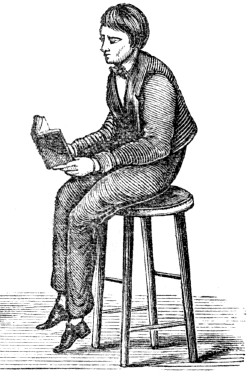
Fig. 29. The position assumed when the seat is of proper height, and the feet supported.
Fig. 30. The position a child naturally assumes when the seat is so high that the feet are not supported.
134. What is said of the benches or chairs in a school-room? What is represented by fig. 29? By fig. 30? What is the effect when the lower limbs are not supported?
135. Compression of the chest should be avoided. In children, and also in adults, the ribs are very flexible, and a small amount of pressure will increase their curvature, particularly at the lower part of the chest, and thus lessen the size of this cavity. The lower ribs are united to the breast-bone, by long, yielding cartilages, and compression may not only contract the chest, but an unseemly and painful ridge may be produced, by the bending of the cartilages, on one or both sides of the sternum.
Fig. 31.
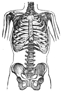
Fig. 32.
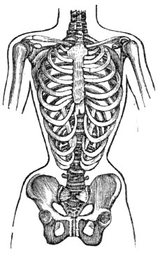
Fig. 31. A natural and well-proportioned chest.
Fig. 32. A chest fashionably deformed.
135. Why should compression of the chest be avoided? What is represented by fig. 31? By fig. 32?
136. Again, the cartilages on one side may be bent outward, while those on the opposite side are bent inward, thus forming a depression parallel with the sternum. In some instances, the anterior extremity of the lower ribs on each side are brought nearly or quite together. In these instances, the movable extremities of the ribs are drawn down toward the haunch-bones, while the space between the ribs is lessened. All this may be effected by tight or “snug” clothing. Therefore the apparel of a child should be loose, and supported over the shoulders, to avoid the before-mentioned evils. The same may be said of the clothing for adults.
137. The erect position in sitting and standing should be assiduously observed. The spinal column, in its natural position, curves from front to back, but not from side to side The admirable arrangement of the bones, alternating with cartilages, permits a great variety of motions and positions; and when the spine is inclined to either side, the elasticity of its cartilages tends to restore it to its natural position. For this reason we may incline the spinal column in any direction for a short time, without danger of permanent curvature, if, afterward, the erect position is assumed.[4]
138. But if a stooping position, or a lateral curved posture, is continued for a long time, the spinal column does not easily recover its proper position, for the compressed edges of the cartilages lose their power of reaction, and finally one side of the cartilage becomes thinned, while the other is thickened; and these wedge-shaped cartilages produce a permanent curvature of the spinal column. In a similar way, the student, seamstress, artisan, and mechanic acquire a stooping position, and become round shouldered, by inclining forward to bring their books or work nearer the eyes.
136. May simply “snug” clothing compress the cartilages? How should the apparel of a child be worn? 137. In what direction does the spinal column, in its natural position, curve? What restores it to its natural position when curved laterally? 138. What is the effect if a lateral curved position of the spinal column is continued for a long time? 139. When one shoulder is elevated for a long time, what is the effect upon the spinal column?
139. Pupils, while writing, drawing, and sometimes while studying, frequently incline the spinal column to one side, in order to accommodate themselves to the desks at which they are seated. Often, these are higher than the elbow as it hangs from the shoulder while at rest. This attitude elevates one shoulder while it depresses the other; consequently, the upper part of the spinal column is inclined toward the elevated shoulder, and the lower part is curved in the opposite direction, giving the form of the letter S to the supporting column of the body.
Fig. 33.
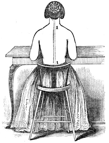
Fig. 33. The table is of proper height, the position is correct, and the spinal column, 1, 1, is straight, while the shoulders are of equal height.
What does fig. 33 represent?
Experiment. Let a pupil be placed at a desk or table with one elbow raised, as is frequently seen while writing, or at study, and observe the condition of the shoulder and spinal column in this position. Place another pupil at a table no higher than the elbow when it hangs by the side while sitting, and observe the appearance of the shoulders and spinal column. By a comparison of the two attitudes, the preceding remarks will be comprehended and appreciated.
Fig. 34.
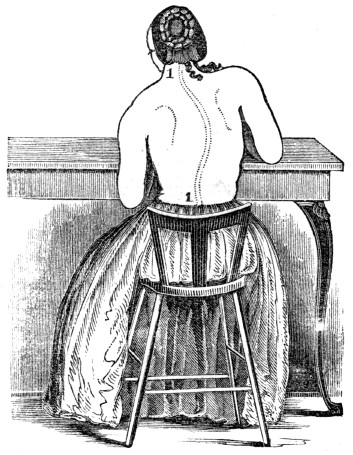
Fig. 34. The table is too high, and the position is oblique and improper. The right shoulder is seen higher than the left, while the spinal column, 1, 1, exhibits three curves.
140. One shoulder may be elevated, and no injurious results follow, provided care is taken not to keep it in the raised position too long, or if the opposite shoulder is elevated for the same period of time. The right shoulder projects more frequently than the left. This arises from the greater use of the right hand with the shoulder elevated, and not unfrequently the oblique positions assumed in performing the daily vocations of life. With proper care, and by calling into action the left shoulder, this deformity can be prevented.
What experiment is mentioned? What does fig. 34 represent? 140. How can one shoulder be elevated and no injurious results follow?
Fig. 35.
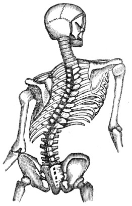
Fig. 35. A representation of a deformed trunk.
141. The loss of symmetry and diminution of height from deformed spines are minor considerations, compared with the distortions that the chest experiences, thereby impairing respiration and inducing diseases of the heart and lungs. The invasion of the functions of these two important organs lessens the vitality of the whole system, and causes general ill health. Again, the curvature of the spinal column is frequently attended by irritation and disease of the spinal cord.
Why does the right shoulder project more frequently than the left? How can this deformity be prevented? 141. What is said of deformed spinal columns?
142. Eminent physicians, both in this country and France state that not more than one female in ten, who has been fashionably educated, is free from deformities of the shoulder or spinal column. Teachers, as well as mothers, should notice the positions of the child in performing the tasks allotted to it, whether studying or pursuing any employment. The feebler the organization of the child, the more frequently should there be a change of position.
143. When a slight projection of the shoulder, with a curvature of the spine, exists, it can be improved by walking with a book, or something heavier, upon the head; to balance which, the spinal column must be nearly erect. Those people that carry burdens upon their heads seldom have crooked spines.
Observation. Persons from the North, in travelling through the Southern States, are surprised to see the heavy burdens that the porters carry on their heads. It is not unusual to see them walking at a rapid pace, with one or two trunks, weighing fifty or eighty pounds each, upon their heads. Occasionally, we meet an itinerant toy-man, with his tray of fragile merchandise upon his head, walking with as much apparent security, as though his toys, or images, were in his hands. This is the easiest method of carrying burdens, because the position of the head and spinal column is erect.
144. If the animal and earthy matter of the bones is not deposited in proper proportions, they are deficient in strength. If the gelatin predominates, the bones are weak, and become distorted. When nutrition is defective in the cylindrical bones, the heads are generally enlarged, and the shafts crooked; if in the spinal column, it may be curved; or in the cranium, it may be enlarged. This disease is familiarly known by the name of rickets. It is most common among these who have poor and insufficient food, live in dark, damp rooms, and breathe a vitiated air. The prevention and remedies for this disease are cleanliness, regular exercise, pure air, and nutritious food.
142. What statement by eminent physicians respecting deformities of the spine? What caution to teachers and mothers? 143. Why should we stand and sit erect? How may slight deformities of the spine be prevented? What is frequently noticed in travelling South? 144. What is the effect upon the bones when the gelatin preponderates?
145. When a bone is broken, some days elapse before the substance that reunites it is thrown out from the blood. In young persons, it may be secreted during the second or third week, and in individuals advanced in life, usually during the third and fourth week. When the bone is uniting, during the second, third, or fourth week, the attention of a surgeon is more needed than during the first week. At this time, the ends of the bone should be placed together with accuracy, which requires the careful application of proper dressing. After the bones have united, it will take some weeks to consolidate the uniting material and render the “callus,” or union, firm. During this time, the limb should be used with care.
Observation. When a bone is fractured, a surgeon is immediately called, and the bone is “set.” While the limb remains swelled and painful, the surgeon is required to attend and keep the dressings (bandages and splints) on. When the swelling has abated, and the pain subsided, frequently the patient intimates to the surgeon that his services can be dispensed with, as the “limb is doing well.” This is the most important period, as the bone is uniting, and, unless the ends are nicely adjusted, the dressing properly applied, the person will find, on recovery, a shortened and crooked limb. The surgeon is then censured, when he is not blamable.
What is one cause of rickets? What are the prevention and remedies for this disease? 145. Does the time vary when the reuniting substance of the bone is secreted from the blood? When is the surgeon’s care most needed? Why?
146. It is seldom that a bone is displaced without injury to the connecting ligaments and membranes. When these connecting bands are lacerated, pain, swelling, and other symptoms indicating inflammation succeed, which should be removed by proper treatment, directed by a surgical adviser.
147. In sprains, but few, if any, of the fibres of the connecting ligaments are lacerated; but they are unduly strained and twisted, which occasions acute pain at the time of the injury. This is followed by inflammation and weakness of the joints. The treatment of these injuries is similar to that of a dislocated bone after its reduction. The most important item in the treatment during the few first days, is rest.
148. In persons of scrofulous constitutions, and those in whom the system is enfeebled by disease, white swellings and other chronic diseases of the joints frequently succeed sprains. Such persons cannot be too assiduous in adopting a proper and early treatment of injured joints.
146. What parts are injured in the displacement of a bone? 147. What causes the acute pain in sprains? What is a good remedy for this kind of injury? 148. What caution to persons of scrofulous constitutions?
CHAPTER VIII
THE MUSCLES.
149. All the great motions of the body are caused by the movement of some of the bones which form the framework of the system; but these, independently of themselves, have not the power of motion, and only change their position through the action of other organs attached to them, which, by contracting, draw the bones after them. In some of the slight movements, as the winking of the eye, no bones are displaced. These moving, contracting organs are the Mus´cles, (lean meat.)
ANATOMY OF THE MUSCLES.
150. The MUSCLES, by their size and number, constitute the great bulk of the body, upon which they bestow form and symmetry. In the limbs, they are situated around the bones, which they invest and defend, while they form, to some of the joints, their principal protection. In the trunk, they are spread out to enclose cavities, and constitute a defensive wall, capable of yielding to internal pressure, and reassuming its original state.
151. In structure, a muscle is composed of fas-cic´u-li (bundles of fibres) of variable size. These are enclosed in a cellular membranous investment, or sheath. Every bundle composed of a number of small fibres, and each fibre consists of a number of filaments, each of which is enclosed in a delicate sheath. Toward the extremity of the organ the muscular fibre ceases, and the cellular structure becomes aggregated, and so modified as to constitute ten´dons, (cords,) by which the muscle is tied to the surface of the bone. The union is so firm, that, under extreme violence, the bone will sooner break than permit the tendon to separate from its attachment. In some situations, there is an expansion of the tendon, in the manner of a membrane, called Ap-o-neu-ro´sis, or Fas´ci-a.
149. How are all the motions of the body produced? What are these motor organs called? 150–160. Give the anatomy of the
muscles.
150. What is said of the muscles? 151. Give their structure.Observation. The pupil can examine a piece of boiled beef, or the leg of a fowl, and see the structure of the fibres and tendons of a muscle.
Fig. 36.
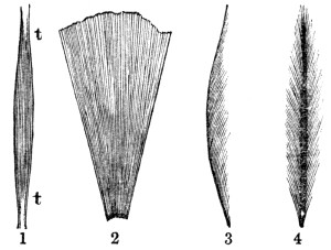
Fig. 36. 1, A representation of the direction and arrangement of the fibres in a fusiform, or spindle-shaped muscle. 2, In a radiated muscle. 3, In a penniform muscle. 4, In a bipenniform muscle. t, t, The tendons of a muscle.
152. Muscles present various modifications in the arrangement of their fibres, as relates to their tendinous structure. Sometimes they are completely longitudinal, and terminate, at each extremity, in a tendon, the entire muscle being spindle-shaped. In other situations, they are disposed like the rays of a fan, converging to a tendinous point, and constituting a ra´di-ate muscle. Again they are pen´ni-form, converging, like the plumes of a pen, to one side of a tendon, which runs the whole length of the muscle; or they are bi-pen´ni-form, converging to both sides of the tendon.
How are tendons or cords formed? What is the expansion of a tendon called? How can the structure of muscles and their fibres be shown? What does fig. 36 represent? 152. Give the different arrangements of muscular fibres.
153. In the description of a muscle, its attachments are expressed by the terms “origin” and “insertion.” The term origin is generally applied to the more fixed or central attachment, or to the point toward which motion is directed; while insertion is assigned to the more movable point, or to that most distant from the centre. The middle, fleshy portion is called the “belly,” or “swell.” The color of a muscle is red in warm-blooded fish and animals; and each fibre is supplied with arteries, veins, lymphatics, and both sensitive and motor nervous filaments.
154. The FASCIA is of various extent and thickness, distributed through the different regions of the body, for the purpose of investing and protecting the softer and more delicate organs. An instance is seen in the membrane which envelopes a leg of beef, and which is observed on the edges of the slices when it is cut for broiling. When freshly exposed, it is brilliant in appearance, tough, and inelastic. In the limbs it forms distinct sheaths to all the muscles.
155. This tendinous membrane assists the muscles in their action, by keeping up a tonic pressure on their surface. It aids materially in the circulation of the fluids, in opposition to the laws of gravity. In the palm of the hand and sole of the foot, it is a powerful protection to the structures that enter into the formation of these parts. In all parts of the system, the separate muscles are not only invested by fascia, but they are arranged in layers, one over another. The sheath of each muscle is loosely connected with another, by the cellular membrane.
153. What is meant by the origin of a muscle? The insertion? The swell? What is the color of muscles? With what is each muscular fibre supplied? 154. What is said of fascia? What is its appearance when freshly exposed? 155. What effect has it on the muscles? Give other uses of the fascia.
156. The interstices between the different muscles are filled with adipose matter, or fat. This is sometimes called the packing of the system. To the presence of this tissue, youth are indebted for the roundness and beauty of their limbs.
Fig. 37.
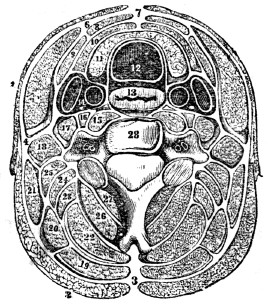
Fig. 37. A transverse section of the neck. The separate muscles, as they are arranged in layers, with their investing fasciæ, are beautifully represented. As the system is symmetrical, figures are placed only on one side. In the trunk the muscles are arranged in layers, surrounded by fasciæ, as in the neck. The same is true of the muscles of the upper and lower limbs.
12, The trachea, (windpipe.) 13, The œsophagus, (gullet.) 14, The carotid artery and jugular vein. 28, One of the bones of the spinal column. The figures that are placed in the white spaces represent some of the fasciæ; the other figures indicate muscles.
157. The muscles may be arranged, in conformity with the general division of the body, into four parts: 1st. Those of the Head and Neck. 2d. Those of the Trunk. 3d. Those of the Upper Extremities. 4th. Those of the Lower Extremities.
156. Give a reason why the limbs of youth are rounder than those of the aged. Describe fig. 37.
Fig. 38.
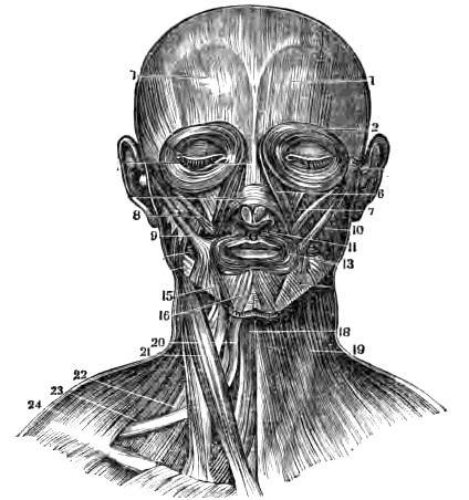
Fig. 38. The superficial layer of muscles on the face and neck. 1, 1, The occipito-frontalis muscle. 2, The orbicularis palpebrarum. 6, The levator labii superioris 7, The levator anguli oris. 8, The zygomaticus minor. 9, The zygomaticus major 10, The masseter. 11, The depressor labii superioris. 13, The orbicularis oris. 15, The depressor anguli oris. 16, The depressor labii inferioris. 18, The sterno-hyoideus. 19, The platysma-myodes. 20, The superior belly of the omo-hyoideus. 21, The sterno-cleido mastoideus. 20, The scalenus medius. 23, The inferior belly of the omo-hyoideus. 24, The trapezius.[5]
Practical Explanation. The muscle 1, 1, elevates the eyebrows. The muscle 2 closes the eye. The muscle 6 elevates the upper lip. The muscles 7, 8, 9, elevate the angle of the mouth. The muscle 10 brings the teeth together when eating. The muscle 11 depresses the upper lip. The muscle 13 closes the mouth. The muscle 15 depresses the angle of the mouth. The muscle 16 draws down the lower lip. The muscles 18, 19, 20, 23, depress the lower jaw and larynx and elevate the sternum. The muscle 21, when both sides contract, draws the head forward, or elevates the sternum; when only one contracts, the face is turned one side toward the opposite shoulder. The muscles 18, 19, 20, 21, 22, 23, 24, aid in respiration.
Observation. When we are sick, and cannot take food, the body is sustained by absorption of the fat. The removal of it into the blood causes the sunken cheek, hollow eye, and prominent appearance of the bones after a severe illness.
158. The number of muscles in the human body is more than five hundred; in general, they form about the skeleton two layers, and are distinguished into superficial and deep-seated muscles. Some of the muscles are voluntary in their motions, or act under the government of the will, as those which move the fingers, limbs, and trunk; while others are involuntary, or act under the impression of their proper stimulants, without the control of the individual, as the heart.
Observations. 1st. The abdominal muscles are expiratory, and the chief agents for expelling the residuum from the rectum, the bile from the gall bladder, the contents of the stomach and bowels when vomiting, and the mucus and irritating substances from the bronchial tubes, trachea, and nasal passages by coughing and sneezing. To produce these effects they all act together. Their violent and continued action sometimes produces hernia, and, when spasmodic, may occasion ruptures of the different organs.
2d. The contraction and relaxation of the abdominal muscles and diaphragm stimulate the stomach, liver, and intestines to a healthy action, and are subservient to the digestive powers. If the contractility of their muscular fibres is destroyed or impaired, the tone of the digestive apparatus will be diminished, as in indigestion and costiveness. This is frequently attended by a displacement of those organs, as they generally gravitate towards the lower portion of the abdominal cavity, when the sustaining muscles lose their tone and become relaxed.
What causes the hollow eye and sunken cheek after a severe sickness? 158. How many muscles in the human system? Into how many layers are they arranged? What is a voluntary muscle? Give examples. What is an involuntary muscle? Mention examples. Give observation 1st, respecting the use of the abdominal muscles? Observation 2d.
Fig. 39.
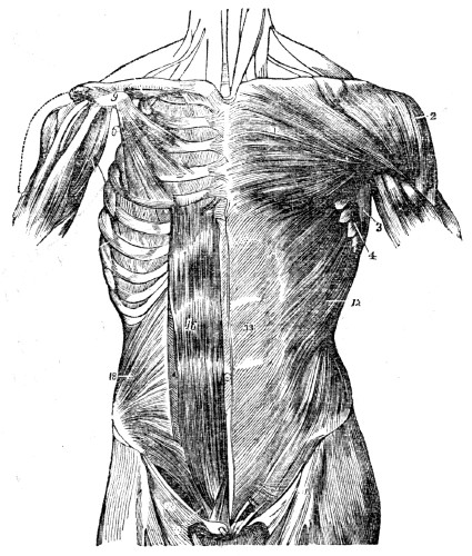
Fig.
39.
A front view of the muscles of the trunk. On the left side thesuperficial
layer is seen; on the right, the deep layer. 1, The pectoralis major muscle. 2, The deltoid muscle. 6, The pectoralis minor muscle. 9, The coracoid process of the scapula. 11, The external intercostal muscle. 12, The external oblique muscle 13, Its aponeurosis. 16, The rectus muscle of the right side. 18, The internal oblique muscle.Practical Explanation. The muscle 1 draws the arm by the side, and across the chest, and likewise draws the scapula forward. The muscle 2 elevates the arm. The muscle 6 elevates the ribs when the scapula is fixed, or draws the scapula forward and downward when the ribs are fixed. The muscles 12, 16, 18, bend the body forward or elevate the hips when the muscles of both sides act. They likewise depress the rib in expiration. When the muscles on only one side act, the body is twisted to the same side.
Explain fig. 39. Give the function of some of the most prominent muscles, from this figure.
[5]
In the plates illustrating the muscular system, the names of such muscles are given as are referred to in the paragraph “Practical Explanation.” These names need not be committed to memory. If a pupil wishes to acquire a knowledge of the general attachment of the muscles represented in the plates, he can do so by comparing the muscular plate with that of the skeleton, (fig. 28.)
[4]
Compare 1, 1, Fig. 28, with 2, 2, 2, Fig. 48.
Fig. 40.
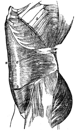
Fig. 40. A lateral view of the muscles of the trunk. 3, The upper part of the external oblique muscle. 4, Two of the external intercostal muscles. 5, Two of the internal intercostals. 6, The transversalis muscle. 7, Its posterior aponeurosis. 8, Its anterior aponeurosis. 11, The right rectus muscle. 13, The crest of the ilium, or haunch-bone.
Practical Explanation. The rectus muscle, 11, bends the thorax upon the abdomen when the lower extremity of the muscle is the fixed point; but when the upper extremity is the fixed point, the effect is to bring forward and raise the pelvis and lower extremities. They likewise depress the ribs in respiration. The transverse muscle, 6, 7, 8, lessens the cavity of the abdomen, and presses the intestines; stomach, and liver upward, against the diaphragm, in expiration.
3d. The region of the back, in consequence of its extent, is common to the neck, the upper extremities, and the abdomen. The muscles of which it is composed are numerous, and are arranged in six layers.
What is represented by fig. 40? Give the function of some of the muscles represented by this figure.
Fig. 41.
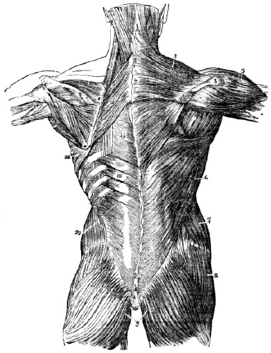
Fig. 41 The first, second, and part of the third layer of muscles of the
back.
The first layer is shown on the right, and the second on the left side. 1, The trapezius muscle. 2, The spinous processes of the vertebræ. 3, The acromion process and spine of the scapula. 4, The latissimus dorsi muscle. 5, The deltoid muscle. 7, The external oblique muscle. 8, The gluteus medius muscle. 9, The gluteus maximus muscle, 11, 12, The rhomboideus major and minor muscles. 15, The vertebral aponeurosis. 16, The serratus posticus inferior muscle. 22, The serratus magnus muscle. 23, The internal oblique muscle.Practical Explanation. The muscles 1, 11, 12, draw the scapula back toward the
spine.
The muscles 11, 12, draw the scapula upward toward the head, and slightly backward. The muscle 4 draws the arm by the side, and backward, The muscle 5 elevates the arm. The muscles 8, 9, extend the thigh on the body. The muscle 1 draws the head back and elevates the chin. The muscle 16 depresses the ribs in expiration. The muscle 22 elevates the ribs in inspiration.159. The diaphragm, or midriff, is the muscular division between the thorax and the abdomen. It is penetrated by the œsophagus on its way to the stomach, by the aorta conveying blood toward the lower extremity, and by the ascending vena cava, or vein, on its way to the heart.
Fig. 42.
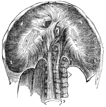
Fig. 42. A representation of the under, or abdominal side of the
diaphragm.
1, 2, 3, 4, The portion which is attached to the margin of the ribs. 8, 10, The two fleshy pillars of the diaphragm, which are attached to the third and fourth lumbar vertebræ. 9, The spinal column. 11, The opening for the passage of the aorta. 12, The opening for the œsophagus. 13, The opening for the ascending vena cava, or vein.Observation. The diaphragm may be compared to an inverted basin, its bottom being turned upward into the thorax, while its edge corresponds with the outline of the edges of the lower ribs and sternum. Its concavity is directed toward the abdomen, and thus, this cavity is very much enlarged at the expense of that of the chest, which is diminished to an equal extent.
159. Describe the diaphragm. What vessels penetrate this muscular septum?
160. “The motions of the fingers do not merely result from the action of the large muscles which lie on the fore-arm, these being concerned more especially in the stronger actions of the hands. The finer and more delicate movements of the fingers are performed by small muscles situated in the palm and between the bones of the hand, and by which the fingers are expanded and moved in all directions with wonderful rapidity.”
Fig. 43.
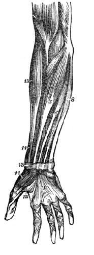
Fig. 44.
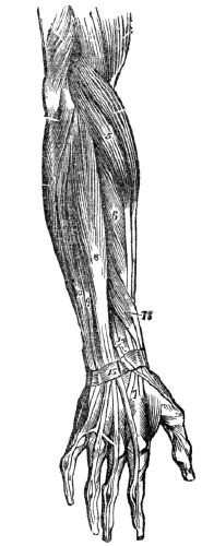
Fig.
43. A front view of the superficial layer of muscles of the fore-arm. 5, The flexor carpi radialis muscle. 6, The palmaris longus muscle. 7, One of the fasciculi of the flexor sublimis digitorum muscle, (the rest of the muscle is seen beneath the tendons of the pintails longus.) 8, The flexor carpi ulnaris muscle. 9, The palmar fascia. 11, The abductor pollicis muscle. 12, One portion of the flexor orevis pollicis muscle. 13, The supinator longus muscle. 14, The extensor ossis metacarpi, and extensor primi internodii pollicis muscles, curving around the lower border of the fore-arm. 15, The anterior portion of the annular ligament, which binds the tendons in their places.Practical Explanation. The muscles 5, 6, 8, bend the wrist on the bones of the fore-arm. The muscle 7 bends the second range of finger-bones on the first. The muscle 11 draws the thumb from the fingers. The muscle 12 flexes the thumb. The muscle 13 turns the palm of the hand upward. The muscles 8, 13, 14, move the hand laterally.
Fig. 44. A back view of the superficial layer of muscles of the fore-arm. 5, The extensor carpi radialis longior muscle. 6, The extensor carpi radialis brevior muscle. 7, The tendons of insertion of these two muscles. 8, The extensor communis digitorum muscle. 9, The extensor minimi dlgiti muscle. 10, The extensor carpi ulnaris muscle. 13, The extensor ossis metacarpi and extensor primi internodii muscles, lying together. 14, The extensor secundi internodii muscle; its tendon is seen crossing the two tendons of the extensor carpi radialis longior and brevior muscles. 15, The posterior annular ligament. The tendons of the common extensor muscle of the fingers are seen on the back of the hand, and their mode of distribution on the back of the fingers.
Practical Explanation. The muscles 5, 6, 10, extend the wrist on the fore-arm. The muscle 8 extends the fingers. The muscle 9 extends the little finger. The muscles 13 extend the metacarpal bone of the thumb, and its first phalanx. The muscle 14 extends the last bone of the thumb. The muscles 10, 13, 14, move the hand laterally.
160. Where are the muscles situated that effect the larger movements of the hand? That perform the delicate movements of the fingers? Give the use of some of the muscles represented by
fig.
43. Those represented by fig. 44.CHAPTER IX.
PHYSIOLOGY OF THE MUSCLES.
161. The muscles exercise great influence upon the system. It is by their contraction that we are enabled to pursue different employments. By their action the farmer cultivates his fields, the mechanic wields his tools, the sportsman pursues his game, the orator gives utterance to his thoughts, the lady sweeps the keys of the piano, and the young are whirled in the mazy dance. As the muscles bear so intimate a relation to the pleasures and employments of man, a knowledge of the laws by which their action is governed, and the conditions upon which their health depends, should be possessed by all.
162. The peculiar characteristic of muscular fibres is contractility, or the power of shortening their substance on the application of stimuli, and again relaxing when the stimulus is withdrawn. This is illustrated in the most common movements of life. Call into action the muscles that elevate the arm, by the influence of the will, or mind, (the common stimulus of the muscles,) and the hand and arm are raised; withdraw this influence by a simple effort of the will, and the muscles, before rigid and tense, become relaxed and yielding.
163. The contractile effect of the muscles, in producing the varied movements of the system, may be seen in the bending of the elbow. The tendon of one extremity of the muscle is attached to the shoulder-bone, which acts as a fixed point; the tendon of the other extremity is attached to one of the bones of the fore-arm. When the swell of the muscle contracts, or shortens, its two extremities approach nearer each other, and by the approximation of the terminal extremities of the muscle, the joint at the elbow bends. On this principle, all the joints of the system are moved. This is illustrated by fig. 45.
161–172. Give the physiology of the muscles. 161. What are some of the influences exerted by the muscles on the system? 162. What is peculiar to muscular fibres? How is this illustrated? 163. Explain how the movements of the system are effected by the contraction of the muscles.
Fig. 45.
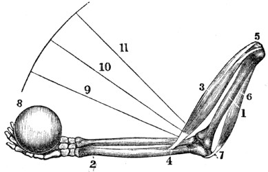
Fig. 45. A representation of the manner in which all of the joints of the body are moved. 1, The bone of the arm above the elbow. 2, One of the bones below the elbow. 3, The muscle that bends the elbow. This muscle is united, by a tendon, to the bone below the elbow, (4,) at the other extremity, to the bone above the elbow, (5,) 6, The muscle that extends the elbow. 7, Its attachment to the point of the elbow. 8, A weight in the hand to be raised. The central part of the muscle 3 contracts, and its two ends are brought nearer together. The bones below the elbow are brought to the lines shown by 9, 10, 11. The weight is raised in the direction of the curved line. When the muscle 6 contracts, the muscle 3 relaxes and the fore-arm is extended.
Experiments. 1st. Clasp the arm midway between the shoulder and elbow, with the thumb and fingers of the opposite hand. When the arm is bent, the inside muscle will become hard and prominent, and its tendon at the elbow rigid, while the muscle on the opposite side will become flaccid. Extend the arm at the elbow, and the outside muscle will swell and become firm, while the inside muscle and its tendon at the elbow will be relaxed.
Explain fig. 45. Give experiment 1st.
2d. Clasp the fore-arm about three inches below the elbow, then open and shut the fingers rapidly, and the swelling and relaxation of the muscles on the opposite sides of the arms, alternating with each other, will be felt, corresponding with the movement of the fingers. While the fingers are bending, the inside muscles swell, and the outside ones become flaccid; and, while the fingers are extending, the inside muscles relax, and the outside ones swell. The alternate swelling and relaxation of antagonist muscles may be felt in the different movements of the limbs.
164. Each fibre of the several muscles receives from the brain, through the nervous filament appropriated to it, a certain influence, called nervous fluid, or stimulus. It is this that induces contraction, while the suspension of this stimulus causes relaxation of the fibres. By this arrangement, the action of the muscular system, both as regards duration and power, is, to a limited extent, under the control of the mind. The more perfect the control, the better the education of the muscular system; as is seen in the graceful, effective, and well-educated movements of musicians, dancers, skaters, &c.
165. The length of time which a muscle may remain contracted, varies. The duration of the contraction of the voluntary muscles, in some measure, is in an inverse ratio to its force. If a muscle has contracted with violence, as when great effort is made to raise a heavy weight, relaxation will follow sooner than when the contraction has been less powerful, as in raising light bodies.
166. The velocity of the muscular contraction depends on the will. Many of the voluntary muscles in man contract with great rapidity, so that he is enabled to utter distinctly fifteen hundred letters in a minute; the pronunciation of each letter requiring both relaxation and contraction of the same muscle, thus making three thousand actions in one minute. But the contraction of the muscles of some of the inferior animals surpasses in rapidity those of man. The race-horse, it is said, has run a mile in a minute; and many birds of prey will probably pass not less than a thousand miles daily.
Give experiment 2d. 164. With what is each muscular fibre supplied? What effect has this stimulus on the muscles? 165. how long does a voluntary muscle remain contracted? 166. On what is the velocity of muscular contraction dependent? How many letters may be pronounced in a minute?
167. The functions of the involuntary muscles are necessary the digestion of food, the absorption and circulation of the nutritive fluids. They could not be trusted with safety to the control of the will, lest the passions or the indiscretions of the person should continually avert those operations so necessary to health, and even to life. The Divine Builder of this complicated machine has wisely ordered that the muscles upon which these motions depend, shall act under the impression of their proper stimulants, without the control of the individual.
168. Again, there are certain operations which could not be safely intrusted to the absolute government of the voluntary muscles, or entirely removed from their control. Thus life can be supported only a few minutes without breathing; but it would be impossible to perform the daily vocations of life if we were compelled to breathe at all times, or at perfectly regular intervals.
169. It has been observed that, among men of the same size, a wide difference exists in their strength and activity—qualities which depend upon the size and number of the nerves, the size and activity of the brain, and the education, or training of the muscles. Men having large nerves leading to the muscles, with the brain active, and muscles well trained will perform feats of strength and agility, that other men, of the same size, cannot effect. Rope-dancers, harlequins, and other performers of feats, are persons thus constituted.
How many contractions and relaxations of the same muscle? What is said of the rapidity of muscular contractions in other animals? 167. When are the involuntary muscles called into action? Why would it not have been safe to trust these important operations to the exclusive control of the will? 168. Give an instance where some of the muscles act under the government of the will, conjoined with those that are involuntary. 169. On what does the difference in muscular activity and strength depend?
170. Persons with small muscles, and largely developed nervous systems, will sometimes exhibit very great muscular power for a time; but it will not be of long continuance, unless the brain is functionally diseased, as in hysteria, delirium of fever, insanity, &c. Men of large muscles and small nerves can never perform feats of great strength; but they have the power of endurance, and are better capacitated for continued labor. Thus we cannot judge of the ability of persons to make exertions and continue them, by their stature alone. Strength, and the power of endurance, are the result of a combination of well-developed muscles, large nerves, and a full-sized, healthy, and active brain.
Observation. The muscles of fishes are large, and the nerves distributed to them, comparatively small. The muscles of birds are small, but their fibres are very compact. The nerves appropriated to the muscles that are called into action in flying, are large as well as numerous.
171. The contractile portion of a muscle is, in general, at a distance from the part to be moved. Thus the principal muscles that move the fingers are situated upon the forearm; and when the limb is nearly or quite extended, the angle formed by the part to be moved and the contractile muscles is small. Again, the attachment of the muscles to the part to be moved is near the joint that forms the fulcrum, (fig. 45.) By these arrangements there is a loss of power; but we are compensated for this disadvantage by increased celerity of movement, beauty of form, and adaptation of the limbs to the varied pursuits of man.
170. What is said of those persons who have small muscles and largely developed nervous systems? Of those who have large muscles and small nerves? Upon what do strength and the power of endurance depend? 171. Why is there a loss of power in the action of the muscles?
Illustration. The muscle that bends the elbow acts at disadvantage, and this is greatest when the arm is nearly or quite extended, as the angle of action is then least. This disadvantage is further increased by the attachment of the motive muscles near the joint.
172. The number of muscles which are called into action in the movements of the different joints, varies. The hinge-joints, as the elbow, have two sets of muscles—one to bend the joint, the other to extend it. The ball and socket joints, as the shoulder, are not limited to mere flexion and
extension.
No joint in the system has the range of movement that is possessed by that of the shoulder. By the action of the muscles attached to the arm, it is not only carried upward and forward, but forward and backward. Hence the arm may be moved at any angle, by a combined action of its muscles.Observation. “Could we behold properly the muscular fibres in operation, nothing, as a mere mechanical exhibition, can be conceived more superb than the intricate and combined actions that must take place during our most common movements. Look at a person running or leaping, or watch the motions of the eye. How rapid, how delicate, how complicated, and yet how accurate, are the motions required! Think of the endurance of such a muscle as the heart, that can contract, with a force equal to sixty pounds, seventy-five times every minute, for eighty years together, without being weary.”
Note. It would be a profitable exercise for pupils to press their fingers upon prominent muscles, and, at the same time, vigorously contract them, not only to learn their situations, but their use; as the one that bends the arm, 14, fig. 46.
How is this illustrated? 172. Do all joints require the same number of muscles, when called into action? How many are called into action in the movement of the elbow? What is their office? What is said of the movement of the ball and socket joint?
Fig 46.
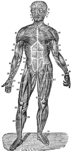
Fig. 46. An anterior view of the muscles of the body. 1. The frontal swell of the occipito-frontalis. 2, The orbicularis palpebrarum. 3, The levator labli superioris. 4, The zygomaticus major. 5, The zygomaticus minor. 6, The masseter. 7, The orbicularis oris. 8, The depressor labli inferioris. 9. The platysma myodes. 10, The deltoid. 11, The pectoralis major. 12, The latissimus dorsi. 14, The biceps flexor cubiti. 15, The triceps extensor cubiti. 16, The supinator radii longus. 18, The flexor carpi radialis longior. 19, The flexor communis digitorum. 20, The annular ligament. 21, The palmar fascia. 22, The obliquus externus abdominis. 26, The psoas magnus. 27, The adductor longus. 28, The sartorius. 29, The rectus femoris. 30, The vastus externus. 31, The vastus internus. 32, The tendon patellæ. 33, The gastrocnemius. 34, The tibialis anticus. 36, The tendons of the extensor digitorum communis.
Fig 47.
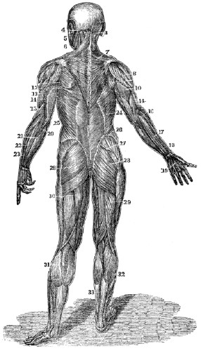
Fig. 47. A posterior view of the muscles of the body. 3, The complexus. 4, The splenius. 5, The masseter. 6, The sterno-cleido mastoideus. 7, The trapezius. 8, The deltoid. 10, The triceps extensor. 13, The tendinous portion of the triceps. 14, The anterior edge of the triceps. 15, The supinator radii longus. 17, The extensor communis digitorum. 18, The extensor ossis metacarpi pollicis. 19, The tendons of the extensor communis digitorum. 20, The olecranon process of the ulna and insertion of the triceps. 21, The extensor carpi ulnaris. 22, The extensor communis digitorum. 24, The latissimus dorsi. 25, Its tendinous origin. 26, The obliquus externus. 27, The gluteus medius. 28, The gluteus magnus. 29, The biceps flexor cruris. 30, The semi-tendinosus. 31, 32, The gastrocnemius. 33, The tendo Achillis.
Practical Explanation. The muscle 1, fig. 46, by its contraction, raises the eyebrows. The muscle 2, fig. 46, closes the eyelids. The muscle 3, fig. 46, elevates the upper lip. The muscles 4, 5, fig. 46, elevate the angles of the mouth. The muscles 6, fig. 46, and 5, fig. 47, bring the teeth together. The muscle 7, fig. 46, closes the mouth. The muscle 8, fig. 46, depresses the lower lip. The muscles 9, fig. 46, and 6,
fig.
47, bend the neck forward. The muscles 3, 4, fig. 47, elevate the head and chin. The muscle 22, fig. 46, bends the body forward, and draws the ribs downward. The muscle 11, fig. 46, brings the shoulder forward. The muscle 7, fig. 47, draws the shoulder back. The muscles 10, fig. 46, and 8, fig. 47, elevate the arm. The muscles 11, fig. 46, and 24, fig. 47, bring the arm to the side. The muscle 14, fig. 46, bends the arm at the elbow. The muscle 10, fig. 47, extends the arm at the elbow. The muscles 16, 18, fig. 46, bend the wrist and fingers. The muscle 19 bends the fingers. The muscles 18, 21, 23, fig. 47, extend the wrist. The muscle 23, fig. 47, extends the fingers. The muscles 26, 27, 28, fig. 46, bend the lower limbs on the body, at thehip.
The muscle 28, fig. 46, draws one leg over the other, (the position of a tailor when sewing.) The muscles 27, 28,fig.
47,
extend the lower limbs on the body, at thehip.
The muscles 29, 30, 31, fig. 46, extend the leg at the knee. The muscles 29, 30, fig.47,
bend the leg at the knee. The muscles 34, 36, fig. 46, bend the foot at the ankle, and extend the toes. The muscles 31, 32, 33, fig. 47, extend the foot at the ankle.Note. Let the anatomy and physiology of the muscular system be reviewed, in form of topics, from figs 46, 47, or from the anatomical outline plates No. 3 and 4.
CHAPTER X.
HYGIENE OF THE MUSCLES
173. The muscles should be used, in order that the size and strength of these organs may be adequate to the demand made upon them. It is a law of the system that the action and power of an organ are commensurate, to a certain extent, with the demand made upon it; and it is a law of the muscular system that, whenever a muscle is called into frequent use, its fibres increase in thickness within certain limits, and become capable of acting with greater force; while, on the contrary, the muscle that is little used decreases in size and power.
Illustrations. 1st. The blacksmith uses and rests the muscles of his arm when striking upon the anvil. They not only increase in size, but become very firm and hard.
2d. The student uses the muscles of the arm but little, in holding his books and pen; they not only become small, but soft.
3d. Let the student leave his books, and wield an iron sledge, and the muscles of his arm will increase in size and firmness. On the other hand, let the blacksmith assume the student’s vocation, and the muscles of his arm will become soft and less firm.
174. When the muscles are called into action, the flow of blood in the arteries and veins is increased. The increased flow of blood in the arteries and veins, causes a more rapid deposition of the particles of matter of which the muscles are composed. If the exercise is adequate to the power of the system, the deposit of new material will exceed in quantity the particles of matter removed, and both the size and energy of the muscles are increased. But there is a limit to the muscles becoming strong by labor. Sooner or later, man will attain his growth or power; yet by judicious exercise, care, and discreet management, the greatest power of the muscles may be preserved until advanced age.
173–211. Give the hygiene of the muscles. 173. What is necessary that muscles may attain size and strength? Give a law of the muscular system. Show this by practical illustrations. 174. Why do muscles increase in size when exercised?
175. The muscles are lessened in size and diminished in power when the exercise is continued so as to produce a feeling of exhaustion. The loss of material, in this instance, will exceed the deposition of the atoms of matter. This is seen in the attenuated frames of over-tasked domestic animals, as the horse. The same truth is illustrated by the laborious agriculturist, who, in consequence of too severe toil while gathering the products of the field, frequently diminishes his weight several pounds in a few weeks. Exercise, either for pleasure or profit, may fatigue, yet it should never be protracted to languor or exhaustion, if the individual desires “a green old age.”
176. The same amount of exercise will not conduce to the health of all individuals. If riding or walking one mile causes slight fatigue, this may be beneficial; while, by travelling two miles, the exhaustion may be highly injurious. Exercise and labor should be adapted to the strength of particular individuals. How little soever the strength, that must be the measure of exertion. Any other rule would be fatal to the hopes of invigorating the system, either by exercise or labor.
Is there a limit to the muscles becoming powerful by action? How may the strength of muscles be kept until advanced age? 175. What is the effect when exercise is continued until there is a feeling of exhaustion? Give a practical illustration. What rule is mentioned in regard to exercise? 176. Can all persons take the same amount of exercise? What rule is given as to the amount of exercise?
177. Relaxation must follow contraction, or, in other words, rest must follow exercise. The necessity of relaxation, when a muscle has been called into action, is seen in the example of a boy extending his arm with a book in his hand, as a penalty. The boy can keep the arm extended but a short time, make what effort he may. It is also seen in the restlessness and feverish excitement that are evinced by persons gazing on troops during days of review. The same is noted in shopping. Such employments call into action the muscles that support the spinal column in an erect position, and the languor or uneasiness is muscular pain. The long-continued tension of a muscle enfeebles its action, and eventually destroys its contractility.
178. In school, the small children, after sitting a short time, become restless. If their position be changed, their imperfectly developed muscles will acquire tone, and will again support the spinal column erect without pain. The necessity for frequent recesses in school, is founded on the organic law of muscular action alternating with rest. The younger and feebler pupils are, the greater the necessity for frequent recesses. We would not have the teacher think that one half of the time should be spent in recesses; or the mother, that her daughter is going to school to play. But we do maintain that recesses should be given, and that they should be short and frequent, especially for small and feeble scholars.
179. Exhaustion is the inevitable result of continued muscular contraction. For example, let a lady ply the needle quickly for some hours, and the muscles of the back and right arm will become exhausted, which will be indicated by a sense of weariness in these parts. A change of employment and position calls into action a different set of muscles, and the exhausted organs are relieved.
177. What is said of the contraction and relaxation of the muscles? Give examples of the necessity of relaxing the muscles. 178. Why should not small children be confined in one position for a long time? What evils result from this practice? What class of pupils should have recesses most
frequently?
179. What effect has continued muscular contraction?180. Much more labor will be accomplished by taking time to relax the exhausted muscles, or by so changing the employment as to bring into action a new set of muscles; the woodman thus relieves himself, by sawing and splitting alternately. This principle applies to the labor of the horse and ox; and it is also applicable to all kinds of employment. With the invalid convalescing from fever, relapses result from inattention to these laws. When a patient is recovering from sickness, his physician should take care that his exercise be proper, neither too much, too little, nor too long continued.
181. The muscles of growing youths will not endure so much exercise or labor as those of mature men. In youth a portion of the vital, or nervous energy of the system, is expended upon the growth of the organs of the body, while in the individual who has attained his growth, this expenditure is not demanded; consequently severe labor or exercise should not be imposed on growing children.
Observation. In the campaigns of Napoleon Bonaparte, his army was frequently recruited by mere boys. He complained to the French government, because he was not supplied with men of mature years, as the youths could not endure the exertion of his forced marches.
182. The muscles should be gradually called into action. These organs in action require more blood and nervous fluid than when at rest. As the circulation of these fluids can only be increased in a gradual manner, it follows, that, when the muscular system has been in a state of rest, it should not suddenly be called into vigorous action. On arising from a bed, lounge, or chair, the first movements of the limbs should be slow, and then gradually increased.
Observation. if a man has a certain amount of work to perform in nine hours, and his muscles have been in a state of rest, he will do it with less fatigue by performing half the amount of the labor in five hours, and the remainder in four hours. The same principle should be regarded in driving horses and other beasts of burden.
180. How can the greatest amount of labor be secured with the least exhaustion to the muscles? 181. Why should not severe labor be imposed on growing children? 182. How should the muscles be called into action?
183. The muscles should be rested gradually, when they have been vigorously used. If a person has been making great muscular exertion in cutting wood, or any other employment, instead of sitting down to rest, he should continue muscular action, for a short time, by some moderate labor or amusement.
184. If the system has been heated by muscular action, and the skin is covered with perspiration, avoid sitting down “to cool” in a current of air; rather, put on more clothing, and continue to exercise moderately. In instances when severe action of the muscles has been endured, bathing and rubbing the skin of the limbs and joints that have been used, are of much importance. The laboring agriculturist and industrious mechanic, by reducing to practice this suggestion, would thus prevent soreness of the muscles, and stiffness of the joints.
185. The muscles should be abundantly supplied with pure blood. This state of the circulating fluid requires a healthy condition of the digestive apparatus, and that the skin should be kept warm by proper clothing, clean by bathing, and be acted upon by pure air and good light; the movements of the ribs and diaphragm should be unrestricted, and the lungs should have ample volume and be supplied with pure air. In all instances, muscular power is greatest when the preceding conditions exist, as the muscles are then stimulated by pure blood; consequently, it is of practical importance to the mechanic, the farmer, the man of leisure, and not less so to the ladies, to observe these conditions, whatever vocation of life they pursue.
183. How should the muscles be rested when they have been vigorously used? 184. What precaution is given when the skin is covered with perspiration? How may soreness of the muscles, consequent upon severe action, be prevented? 185. Should the muscles be supplied with pure blood? When is muscular power the greatest?
186. The muscles should be used in pure air. The purer the air we breathe, the more stimulating the blood supplied to the muscles, and the longer they can be used in labor, walking, or sitting, without fatigue and injury; hence the benefit derived in thoroughly ventilating all inhabited rooms. For the same reason, if the air of the sick-room is pure, the patient will sit up longer than when the air is impure.
Observation. It is a common remark that sick persons will sit up longer when riding in a carriage, than in an easy chair in the room where they have lain sick. In the one instance, they breathe pure air; in the other, usually, a confined, impure air.
187. The muscles should be exercised in the light. Light, particularly that of the sun, exercises more or less influence on man and the inferior animals as well as on plants. Both require the stimulus of this agent. Shops occupied by mechanics, kitchens, and sitting-rooms, should be well lighted, and situated on the sunny side of the house. Cellar kitchens and underground shops should be avoided. For similar reasons, students should take their exercise during the day, rather than in the evening, and, as much as possible, laborers should avoid night toil.
Illustrations. Plants that grow in the shade, as under trees, or in a dark cellar, are of lighter color and feebler than those that are exposed to the light of the sun. Persons that dwell in dark rooms are paler and less vigorous than those who inhabit apartments well lighted, and exposed to the rays of the sun.
186. Why should the muscles be used in pure air? Give a common observation. 187. What effect has light on the muscular system? What should the laborer avoid? Why should not students take their daily exercise in the evening? How is the influence of solar light illustrated?
188. Exercise should be regular and frequent. The system needs this means of invigoration as regularly as it does new supplies of food. It is no more correct that we devote several days to a proper action of the muscles, and then spend one day inactively, than it is to take a proper amount of food for several days, and then withdraw this supply for a day. The industrious mechanic and the studious minister suffer as surely from undue confinement as the improvident and indolent. The evil consequences of neglect of exercise are gradual, and steal slowly upon an individual. But sooner or later they are manifested in muscular weakness, dyspepsia, and nervous irritability.
Observation. The custom among farmers of enduring severe and undue toil for several successive days, and then spending one or two days in idleness to rest, is injudicious. It would be far better to do less in a day, and continue the labor through the period devoted to idleness, and then no rest will be demanded.
189. Every part of the muscular system should have its appropriate share of exercise. Some employments call into exercise the muscles of the upper limbs, as shoe-making; others, the muscles of the lower limbs; while some, the muscles of both upper and lower limbs, with those of the trunk, as farming. In some kinds of exercise, the lower limbs are mainly used, as in walking; in others, the upper limbs; and again, the muscles of the trunk, together with those of the upper and lower limbs, as in archery, quoits, playing ball. Those trades and kinds of exercise are most salutary, in which all the muscles have their due proportion of action, as this tends to develop and strengthen them equally. Thus labor upon the farm and domestic employment are superior as vocations, and archery, quoits, and dancing, if the air is pure, among the pastimes. For sedentary persons, that kind of exercise is best which calls into action the greatest number of muscles.
188. How should exercise be taken? What is said respecting irregular exercise? Are the consequences of neglected exercise immediately apparent? What practical observation is given? 189. Should every muscle have its due amount of exercise? Mention some employments that only call into action the muscles of the upper limbs. Those of the lower limbs, those of the trunk and limbs. Mention, in the different pastimes, what muscles are called into action.
190. The proper time for labor or exercise should be observed. This is modified by many circumstances. As a general rule, the morning, when the air is pure and the ground dry, is better than the evening; for then, the powers of the body are greatest. Severe exercise and labor should be avoided immediately before or after eating a full meal, for the energies of the system are then required to perform the digestive function. For similar reasons, it is not an appropriate time for energetic muscular action immediately before or after severe mental toil, as the powers of the system are then concentrated upon the brain.[6]
What kinds of exercise are best? 190. What rule is given respecting the time for exercise? 191. Why do the muscles require sleep? What is the effect of an inversion of the law of rest?
191. The muscles require sleep to restore their expended energies. Among the arrangements of creative wisdom, no one harmonizes with the wants of the system more than the alternation of day and night. The natural inclination of man to sleep, is in the stilly hour of night, when all nature reposes, and to be in action during the light of day. An inversion of this law of rest causes greater exhaustion of the system than the same amount of exertion during daylight. This is illustrated by the wearied and exhausted condition of watchers, night-police, and other individuals who spend a part of the night in some active business of life.
192. The muscles should not be compressed. Compression prevents the blood from passing to the muscles with freedom; consequently, they are not supplied with material to renovate and promote their growth. Again, pressure stimulates the lymphatics to action; and by the increased activity of these vessels the muscles are attenuated. In the case of a man with a fractured limb, the muscles are not only enfeebled by inaction, but diminished in size by compression from the dressing. Limbs enfeebled in this way will not recover their size, tone, and strength, until the bandages are removed, and a proper amount of exercise taken.
193. The pressure of tight dresses, under the name of a “snug fit,” enfeebles the muscles of the back, and is a common cause of projecting shoulders and curvature of the spinal column. Thus every appendage to the dress of ladies which prevents free motion of the muscles of the chest and spinal column, weakens the muscles thus restrained, and not only prevents the proper expansion of the lungs, but, by weakening the muscles which sustain the spine, induces curvature and disease. Whalebone, wood, steel, and every other unyielding substance, should be banished from the toilet, as enemies of the human race.
194. The mind exerts a great influence upon the tone and contractile energy of the muscular system. A person acting under a healthy mental stimulus will make exertion with less fatigue than he would without this incentive. For this reason, a sportsman will pursue his game miles without fatigue, while his attendant, not having any mental stimulus, will become weary. Again, if the sportsman spends some hours in pursuit of his favorite game without success, a feeling of languor creeps over him; but while he is thus fatigued and dispirited, let him catch a glimpse of the game,—his wearied feelings are immediately dissipated, and he presses on with renewed energy and recruited strength.
192. Why should not the muscles be compressed? 193. What is the effect of tight clothing upon the muscles? 194. What is said of the influence of the mind upon muscular activity? Give an illustration of mental stimulus coöperating with muscular activity in the case of a sportsman.
195. This principle was well illustrated in the retreat from Russia of the defeated and dispirited French army. When no enemy was near, they had hardly strength sufficient to carry their arms; but no sooner did they hear the report of the Russian guns, than new life seemed to pervade them, and they wielded their weapons powerfully until the foe was repulsed, then there was a relapse to weakness, and prostration followed. It is thus with the invalid when riding for his health;—relate an anecdote, or excite this mental stimulus by agreeable conversation, and much benefit will accrue from the ride to the debilitated person. So it is in the daily vocations of life; if the mind have some incentive, the tiresomeness of labor will be greatly diminished. Let an air of cheerfulness ever pervade our every employment, and, like music, “it sweetens toil.”
196. Facts illustrative of the inutility of calling the muscles into action, without the coöperation of the mind, are seen in the spiritless aspect of many of our boarding school processions, when a walk is taken merely for exercise, without having in view any attainable object. But present to the mind a botanical or geological excursion, and the saunter will be exchanged for the elastic step, the inanimate appearance for the bright eye and glowing cheek. The difference is, simply, that, in the former case, the muscles are obliged to work without that full nervous impulse so essential to their energetic action; and that, in the latter, the nervous influence is in full and harmonious operation.
195. Give an illustration of mental stimulus coöperating with muscular activity in the case of the dispirited French army in their retreat from Russia. How can a union of mental impulse and muscular action be beneficial to an invalid? Does this same principle apply to those who labor? 196. Give an instance of the different effects produced by the absence and presence of the mental
stimulus.
197. It must not, however, be supposed that a walk simply for the sake of exercise can never be beneficial. Every one, unless prevented by disease, should consider it a duty to take exercise every day in the open air; if possible, let it be had in combination with harmonious mental exhilaration; if not, let a walk, in an erect position, be made so brisk as to produce rapid respiration and circulation of the blood, and in a dress that shall not interfere with free motions of the arms and free expansion of the chest.
Observation. The advantages of combining harmonious mental excitement, with muscular activity, is thus given by Dr. Armstrong:—
“In whate’er you sweat,
Indulge your taste. Some love the manly toils
The tennis some, and some the graceful dance;
Others, more hardy, range the purple heath
Or naked stubble, where, from field to field,
The sounding covies urge their lab’ring flight,
Eager amid the rising cloud to pour
The gun’s unerring thunder; and there are
Whom still the mead of the green archer charm.
He chooses best whose labor entertains
His vacant fancy most; the toil you hate
Fatigues you soon, and scarce improves your limbs.”
197. May not a walk, simply as an exercise, be beneficial? What is preferred?
[6]
It appears to be a fact, that no two important organs can be called into intense action at the same time, without injury to both, as well as to the general system. This arises from the circumstance that an organ, when in functional action, attracts fluids (sanguineous and nervous) from other organs of the system. Except in a few instances of high health in youth, the power of the system is not adequate to supply more than one organ in action with the appropriate fluids at the same time.
CHAPTER XI.
HYGIENE OF THE MUSCLES, CONTINUED.
198. The erect attitude lessens the exhaustion of the muscles. A person whose position is erect will stand longer, walk further, and perform more labor, than an individual whose position is stooping, but equal in all other respects. The manly port in an erect attitude, depends chiefly upon the action of the muscles of the back; and it follows that the fewer the muscles in a state of tension, the less the draught upon the nervous system, and the less its exhaustion. Another advantage which attends the erect position is, the trunk and head are balanced upon the bones and cartilages of the spinal column. If the body slightly incline forward, the muscles attached to the posterior side of the spine, by a gentle contraction, will bring it to the perpendicular, and even incline it backward. This is immediately removed by a slight contraction of the muscles upon the anterior side of the spinal column.
199. In the erect position, there is a constant slight oscillation of the body backward and forward, like the movement of a pendulum; while, in the stooping posture, the muscles on the posterior side of the spinal column are kept in a state of continued tension and contraction, to prevent the body from falling forward. This enfeebles the muscles of the back, and exhausts the nervous energy, while the erect position favors their development and power, because there is an alternate contraction and relaxation of the muscles. Again, in the stooping position, the lower limbs are curved at the knee. In this attitude, there is a constant tension of the muscles of the lower extremities, which produces muscular exhaustion.
198. Why will a person who stands erect walk further, and perform more labor, than if he assumed the stooping posture? 199. Why are the muscles of the back so soon exhausted in the stooping position?
Fig. 48.
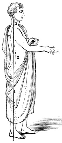
Fig. 49.
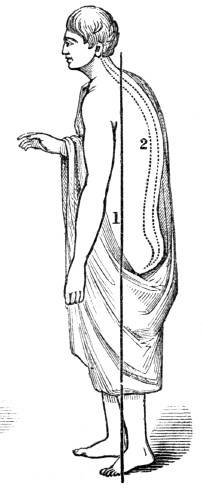
Fig. 48. 1, A perpendicular line from the centre of the feet to the upper extremity of the spinal column, where the head rests. 2, 2, 2, The spinal column, with its three natural curves. Here the head and body are balanced upon the spinal column and joints of the lower extremities, so that the muscles are not kept in a state of tension. This erect position of the body and head is always accompanied with straight lower limbs.
Fig. 49. 1, A perpendicular line from the centre of the feet. 2, Represents the unnatural curved spinal column, and its relative position to the perpendicular,
1.
The lower limbs are curved at the knee, and the body is stooping forward. While standing in this position, the muscles of the lower limbs and back are in continued tension, which exhausts and weakens them.What is represented by figs. 48 and 49?
200. When it is necessary to call into action a part of the muscles of the system in the performance of any duty, as those of the lower limbs in walking, if the muscles of other parts are in a state of inaction, the influence of the nervous system can be determined in an undivided manner upon those parts of the lower limbs in action; hence they will not so soon become wearied or exhausted, as when this influence is divided between a greater number of muscles. In performing any labor, as in speaking, reading, singing, mowing, sewing,
&c.,
there will be less exhaustion, and the effort can be longer maintained in the erect position of the body and head, than in a stooping attitude.Experiment. Hold in each hand a pail of water or equal weights, in a stooping posture, as long as it can be done without much suffering and injury. Again, when the muscular pain has ceased, hold the same pails of water, for the same length of time, in an erect posture, and note the difference in the fatigue of the muscles.
201. If the stooping posture is acquired in youth, we are quite certain of seeing the deformed shoulders in old age. Hence the importance of duly exercising the muscles of the back, for when they are properly developed, the child can and will stand erect. In this attitude, the shoulders will be thrown back, and the chest will become broad and full.
202. Pupils, while standing during recitations, often inadvertently assume the attitude represented by fig. 49, and it is the duty of teachers to correct this position when assumed. When a child or adult has contracted a habit of stooping, and has become round-shouldered, it can be measurably, and generally, wholly, remedied by moderate and repeated efforts to bring the shoulders back, and the spinal column in an erect position. This deformity can and should be remedied in our schools. It may take months to accomplish the desired end, yet it can be done as well under the direction of the kind instructor, as under the stern, military drill sergeant, who never fails to correct this deformity among his raw recruits.
200. What suggestion when it is necessary to call into action a part of the muscular system? Give the experiment that illustrates this principle. 201. Why should a child he taught to stand erect? 202. How can round shoulders acquired by habit be remedied?
Fig. 50.
Fig. 50. A proper position in sitting.
203. The child should be taught to sit erect when employed in study or work. This attitude favors a healthy action of the various organs of the system, and conduces to beauty and symmetry of form. Scholars are more or less inclined to lean forward and place the elbow on the table or desk, for support and this is often done when their seats are provided with backs. Where there is a predisposition to curvature of the spine, no position is more unfavorable or more productive of deformities than this; for it is usually continued in one direction, and the apparent deformity it induces is a projection of the shoulders. If the girl is so feeble that she cannot sit erect, as represented by fig. 50, let her stand or recline on a couch; either is preferable to the position represented by fig. 51. In furnishing school-rooms, care should be taken that the desks are not so low as to compel the pupils to lean forward in examining their books.
203. Why should the erect attitude be assumed in sitting?
Fig. 51.
Fig. 51. An improper position in
sitting.
204. The muscles, when exhausted, cannot endure continued effort. When the energies of the muscular system have been expended by severe and long-continued exercise, or the brain and nervous system prostrated by protracted mental effort, the muscles are unfitted to maintain the body erect in standing or sitting for a long time, as the nervous system, in its exhausted state, cannot supply a sufficient amount of its peculiar influence to maintain the supporting muscles of the body and head in a state of contraction. Hence, a child or adult, when much fatigued, should not be compelled to stand or sit erect in one posture, but should be permitted to vary the position frequently, as this rests and recruits both the muscular and the nervous system.
205. A slight relaxation of the muscles tends to prevent their exhaustion. In walking, dancing, and most of the mechanical employments, there will be less fatigue, and the movements will be more graceful, when the muscles are slightly relaxed. When riding in cars or coaches, the system does not suffer so severely from the jar if there is a slight relaxation of the muscles, as when they are in a state of rigid contraction.
Experiments. Attempt to bow with the muscles of the limbs and trunk rigid, and there will be a stiff bending of the body only at the hip-joint. On the other hand, attempt to bow with the muscles moderately relaxed; the ankle, the knee, and the hip-joint will slightly bend, accompanied with an easy and graceful curve of the body.
206. The muscles when relaxed, together with the yielding character of the cartilage, and the porous structure of the ends of the bones that form a joint, diffuse or deaden the force of jars, or shocks, in stepping suddenly down stairs, or in falling from moderate heights. Hence, in jumping or falling from a carriage, or any height, the shock to the organs of the system may be obviated in the three following ways: 1st. Let the muscles be relaxed, not rigid. 2d. Let the limbs be bent at the ankle, knee, and hips; the head should be thrown slightly forward, with the trunk a little stooping. 3d. Fall upon the toes, not the heel.
204. When are the muscles unfitted to maintain the system erect either in standing or sitting? What is necessary when this condition of the system exists? 205. Why should the muscular system be slightly relaxed in walking, &c.? Give illustrative experiments. 206. What is the reason that we do not feel the jar in falling from a moderate height?
Experiments. Stand with the trunk and lower limbs firm, and the muscles rigid; then jump a few inches perpendicularly to the floor, and fall upon the heels. Again, slightly bend the limbs, jump a few inches, and fall upon the toes, and the difference in the force of the shock, to the brain and other organs, will be readily noticed.
207. The muscles require to be educated, or trained. The power of giving different intonations in reading, speaking, singing, the varied and rapid executions in penmanship, and all mechanical or agricultural employments, depend, in a measure, upon the education of the muscles. In the first effort of muscular education, the contractions of the muscular fibres are irregular and feeble, as may be seen when the child begins to walk, or in the first efforts of penmanship.
208. Repetition of muscular action is necessary. To render the action of the muscles complete and effective, they must be called into action repeatedly and at proper intervals. This education must be continued until not only each muscle, but every fibre of the muscle, is fully under the control of the will. In this way persons become skilful in every employment. In training the muscles for effective action, it is very important that correct movements be adopted at the commencement. If this is neglected, the motions will be constrained and improper, while power and skill will be lost.
How is this shown by experiment? 207. Upon what do the different intonations of sound or mechanical employments depend? Why are the first efforts in educating the muscles indifferent or irregular? 208. Why is repetition of muscular action necessary? Why is it important that correct movements be adopted in the first efforts of muscular education?
Illustration. If a boy, while learning to mow, is allowed to swing his scythe in a stooping position, twisting his body at every sweep of the scythe, he will never become an easy, efficient mower. Proper instruction is as necessary in many of the agricultural branches as in the varied mechanical employments.
Fig. 52.
Fig. 53.
Fig. 52. An improper, but not an unusual position, when writing.
Fig. 53. A proper position, when writing.
209. Good penmanship requires properly trained muscles. To a deficient analysis of the movements of the arm, hand, and fingers, on the part of teachers and pupils in penmanship, together with an improper position in sitting, is to be ascribed the great want of success in acquiring this art. The pen should be held loosely, and when the proper position is attained, the scholar should make an effort to imitate some definite copy as nearly as possible. The movements of the fingers, hand, and arm, necessary to accomplish this, should be made with ease and rapidity, striving, at each effort, to imitate the copy more nearly.
How is this illustrated? 209. Why have so many pupils failed in acquiring good penmanship?
210. When the arm, hand, and fingers are rigid, the large muscles, that bend and extend these parts, are called into too intense action. This requires of the small muscles, that produce the lateral movements, which are essential to rapidity in writing, an effort which they cannot make, or can with difficulty accomplish.
Experiment. Vigorously extend the fingers by a violent and rigid contraction of the muscles upon the lower part of the arm, and the lateral movement which is seen in their separation cannot be made. But gently extend the fingers, and their oblique movements are made with freedom.
211. An individual who is acquainted with the laws of health, whose muscles are well trained, will perform a certain amount of labor with less fatigue and waste to the system, than one who is ignorant of the principles of hygiene, and whose muscles are imperfectly trained. Hence the laboring poor have a deep interest in acquiring a knowledge of practical physiology, as well as skill in their trade or vocation. It is emphatically true to those who earn their bread by the “sweat of their brow,” that “knowledge is power.”
210. What is said of the lateral and oblique movements of the
arm,
hand, and fingers in writing? How is this shown by experiment? 211. Why is the study of physiology and hygiene of utility to the laborer?CHAPTER XII.
THE TEETH.
212. The teeth, in composition, nutrition, and growth, are different from other bones of the body. They vary in number at different periods of life, and, unlike other bones, they are exposed to the immediate action of atmospheric air and foreign substances. The bones of the system, generally, when fractured, unite; but there is never a permanent union of a tooth when broken.
ANATOMY OF THE TEETH.
213. The TEETH are attached to the upper and lower jaw-bone, by means of bony sockets, called al´ve-o-lar processes. These give great solidity to the attachment of the teeth, and frequently render their extraction difficult. The gums, by their fibrous, fleshy structure, serve to fix the teeth more firmly in the jaw.
Observation. When a permanent tooth is extracted, these bony processes are gradually absorbed, so that in advanced age there remains only the jaw-bone covered by the lining membrane of the gum. This accounts for the narrow jaw and falling in of the lips in old age. Frequently, a piece of the alveolar process comes out with the tooth when extracted, and the dentist has then the credit of “breaking the jaw.” No great injury results from the removal of the process in this manner.
212. What is said of the teeth? In what respect do they differ from other bones of the body? 213–218. Give the anatomy of the teeth. 213. What confines the teeth in the jaw-bone? What becomes of the socket when a tooth is removed? What effect has this absorption upon the jaw and lips?
214. The teeth are formed in the interior of the jaws, and within dent´al cap´sules, (membranous pouches,) which are enclosed within the substance of the bone, and present in their interior a fleshy bud, or granule, from the surface of which exudes the ivory, or the bony part of the tooth. In proportion as the tooth is formed, it rises in the socket, which is developed simultaneously with the tooth, and passes through the gum, and shows itself without.
Fig. 54.
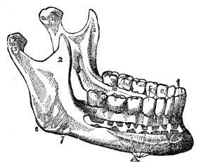
Fig. 54. 1, The body of the lower jaw. 2, Ramus, or branch of the jaw, to which the muscles that move it are attached. 3, 3, The processes which unite the lower jaw with the head. i, The middle and lateral incisor tooth of one side. b, The bicuspid teeth. c, The cuspids, or eye teeth. m, The three molar teeth. A, shows the relation of the permanent to the temporary teeth.
215. The first set, which appears in infancy, is called tem´po-ra-ry, or milk teeth. They are twenty in number; ten in each jaw. Between six and fourteen years of age, the temporary teeth are removed, and the second set appears, called per´ma-nent teeth. They number thirty-two, sixteen in each jaw.
214. Where and how are the teeth formed? Explain fig. 54. 215. What are the first set called? How many in each jaw? The second set? How many in number?
216. The four front teeth in each jaw are called in-ci´sors, (cutting teeth;) the next tooth in each side, the cus´pid, (eye tooth;) the next two, bi-cus´pids, (small grinders;) the next two, mo´lars, (grinders.) The last one on each side of the jaw is called a wisdom tooth, because it does not appear until a person is about twenty years old. The incisors, cuspids, and bicuspids, have each but one root. The molars of the upper jaw have three roots, while those of the lower jaw have but two.
Fig. 55.
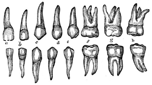
Fig. 55. The permanent teeth of the upper and lower jaw. a, b, The incisors. c, The cuspids. d, e, The bicuspids. f, g, The molars, (double teeth.) h, The wisdom
teeth.
Observation. The shape of the teeth in different species of animals is adapted to the kind of food on which they subsist. Those animals that feed exclusively on flesh, as the lion, have the cuspids, or canine teeth, largely developed, and the molars have sharp cutting points. Those animals that feed on grass and grain, as the horse and the sheep, have their molar teeth more rounded and flat on the crown. The human teeth are adapted to feed on fruits, grain, or flesh, as they are less pointed than those of the cat, and more pointed than those of the sheep.
216. Give the names of the permanent teeth. What teeth have but one root, or “fang”? How many roots have the molars of the upper jaw? Of the lower jaw? What is said of the shape of the teeth in different species of animals?
217. The teeth are composed principally of two substances—the i´vo-ry and the en-am´el. The internal part of the tooth or the ivory, is harder and more enduring than bone, and forms the body of the tooth. The enamel is remarkable for its hardness, and varies somewhat in color with the age, temperament, habits, and manner of living of different individuals. When any part of the enamel is destroyed, it is never regenerated.
Fig. 56.
Fig. 57.
Fig.
56. A side view of the body and enamel of a front tooth.Fig.
57. A side view of a molar tooth. 1, The enamel. 2, The body of thetooth.
3, The cavity in the crown of the tooth that contains the pulp. 4, A nerve that spreads in the pulp of the tooth. 5, An artery that ramifies in the pulp of the tooth.218. Each tooth is divided into two parts, namely, crown and root. The crown is that part which protrudes from the jaw-bone and gum, and is covered by the highly polished enamel. The root, or “fang,” is placed in the sockets of the jaw, and consists of bony matter. Through this bony substance several small vessels pass, to aid in the growth and also in the removal of the tooth. There are, beside these vessels, small white cords passing to each tooth, called nerves. (See fig. 57.) When these nerves are diseased, we have the toothache.
217. Give the structure of the teeth. What is said of the enamel? 218. Into how many parts are the teeth divided? Describe the crown. The root. What vessels pass through the bony matter? What is their use?
PHYSIOLOGY OF THE TEETH.
219. The use of the teeth is twofold. 1st. By the action of the incisors the food is divided, while the molars grind or break down the more solid portions of it. By these processes, the food is prepared to pass more easily and rapidly into the stomach.
220. In the mastication of food there are two movements of the lower jaw—the action by which the teeth are brought together, and the lateral motion. In the former, the food is cut or divided, the jaws acting like shears. This movement is produced by the action of two large muscles situated on each side of the head and face.
Observation. The muscles attached to the lower jaw are of great strength; by their action alone, some persons are enabled to bite the hardest substances. By putting the fingers upon the side of the head above and in front of the ears, and upon the face above the angle of the jaw, while masticating food, the alternate swelling and relaxation of these muscles will be clearly felt.
221. The lateral, or grinding movement of the teeth, is produced by the action of a strong muscle that is attached to the lower jaw on the inside.
Observation. Those animals that live solely on flesh, have only the cutting, or shear-like movement of the jaws. Those that use vegetables for food, have the grinding motion; while man has both the cutting and grinding movement.
219–222. Give the physiology of the teeth. 219. Give one of the functions of the teeth. 220. How many movements of the lower jaw in masticating food? What effect has the first movement upon the food? How produced? What is the character of the masticating muscles? 221. How is the grinding motion of the teeth produced? What is said of the movements of the teeth in different animals?
222. 2d. The teeth aid us in articulating with distinctness certain letters and words. An individual who has lost his front teeth cannot enunciate distinctly certain letters called dental. Again, as the alveolar processes are removed by absorption soon after the removal of the teeth, the lips and cheeks do not retain their former full position, thus marring, in no slight degree, the symmetry of the lower part of the face. Consequently, those simple observances that tend to the preservation of the teeth are of great practical interest to all persons.
HYGIENE OF THE TEETH.
223. To preserve the teeth, they must be kept clean. After eating food, they should be cleansed with a brush and water, or rubbed with a piece of soft flannel, to prevent the tartar collecting, and to remove the pieces of food that may have lodged between them. Toothpicks may be useful in removing any particles inaccessible to the brush. They may be made of bone, ivory, or the common goose-quill. Metallic toothpicks should not be used, as they injure the enamel.
224. The mouth should be cleansed with pure tepid water at night, as well as in the morning; after which the teeth should be brushed upward and downward, both on the posterior and anterior surfaces. It may be beneficial to use refined soap, once or twice every week, to remove any corroding substance that may exist around the teeth; care being taken to thoroughly rinse the mouth after its use.
225. Food or drink should not be taken into the mouth when very hot or very cold. Sudden changes of temperature will crack the enamel, and finally produce decayed teeth.
222. What is another use of the teeth? 223–232. Give the hygiene of the teeth. 223. How can the teeth be preserved? By what means? 224. How often should they be cleansed? 225. What is said of very hot or cold drinks?
Observation. On this account, smoking is pernicious, because the teeth are subjected to an alternate inhalation of both cold and warm air.
226. The temporary teeth should be removed as soon as they become loose. If a permanent tooth makes it appearance before the first is removed, or has become loose, the milk tooth, although not loose, should be removed without delay. This is necessary that the second set of teeth may present a regular and beautiful appearance.
227. In general, when the permanent teeth are irregular, one or more should be removed. If the teeth are crowded and irregular, in consequence of the jaw being narrow and short, or when they press so hard upon each other as to injure the enamel, remove one or more to prevent their looking unsightly, and in a few months the remaining teeth, with a little care, will fill the spaces.
Observation. When it is necessary to remove a tooth, apply to some skilful operator. It requires as much skill and knowledge to extract teeth well, as it does to amputate a limb; yet some persons, who possess strong arms, will obtain a pair of forceps, or a tooth-key, and hang out the sign of “surgeon-dentist,” although ignorant of the principles that should guide them.
228. It is not always necessary to have teeth extracted when they ache. The nerve, or the investing membrane of the root, may be diseased, and the tooth still be sound. In such instances, the tooth should not be extracted, but the diseased condition may be remedied by proper medication. There are many sound teeth, that become painful, as already mentioned, which are unnecessarily removed.
Why is smoking injurious to the teeth? 226. What remark respecting the temporary teeth? 227. What remarks respecting the permanent teeth? Do those persons that extract teeth require skill as well as knowledge? 228. Why should not teeth be extracted at all times when they are painful?
Illustration. Dr. H. M., of Belfast, Me., related to me that an individual in that vicinity had his teeth, (all of them sound,) on one side of the lower jaw, extracted by an ignoramus of a “tooth-puller,” and this without any relief from pain. The disease was tic douloureux, which was relieved by Dr. M.
229. The preservation of the teeth requires that they be frequently examined. When a part of the enamel is removed, and a small portion of the body of the tooth has become carious, in many instances such teeth may be preserved from further decay by having them filled or “plugged” with gold foil. All amalgams, pastes, and cheap patent articles for filling, should be avoided, if you would preserve both the teeth and the general health.
230. The practice of cracking nuts with the teeth, or of lifting heavy bodies, and the constant habit of biting thread, should be avoided, as they finally destroy the enamel.
231. All acidulated drinks and mineral waters, that “set the teeth on edge,” are injurious. All tooth-powders and washes that contain any article that is acid, corrosive, or grinding, should be banished from the toilet. Tobacco is not a preservative of the teeth. It contains “grit,” which wears away the enamel; beside, when chewed, it debilitates the vessels of the gums, turns the teeth yellow, and renders the breath and the appearance of the mouth disagreeable.
232. Healthy persons have generally sound teeth, while feeble persons have decayed teeth. For this reason, we should try to learn and practise the few simple rules that promote health.
Give an illustration of the removal of sound teeth. 229. How may decaying teeth be preserved? What should be avoided in the filling of teeth? 230. What practices should also be avoided? 231. What is said of acidulated drinks? What effect has the chewing of tobacco upon the teeth? 232. What is one reason for preserving health?
CHAPTER XIII.
THE DIGESTIVE ORGANS.
233. From the earliest existence of the human system to the last ray of life, change is impressed upon it by the Giver of this curious fabric. New atoms of matter are deposited, while the old and now useless particles are constantly removed. The material necessary to sustain the growth of the body in early life, and also to repair the waste that is unceasing to animal existence, is the food we eat.
234. Food, animal or vegetable, contains most of the elements of the different tissues of the system, yet it must undergo certain essential alterations before it can become a part of the body. The first change is effected by the action of the Digestive Organs.
ANATOMY OF THE DIGESTIVE ORGANS
235. The DIGESTIVE ORGANS are the Mouth, Teeth,[7] Sal´i-va-ry Glands, Phar´ynx, Œ-soph´a-gus, (gullet,) Stom´ach, In-tes´tines, (bowels,) Lac´te-als, (milk, or chyle vessels,) Tho-rac´ic Duct, Liv´er, and the Pan´cre-as, (sweetbread.)
233. What is impressed upon the human system from its earliest existence? What maintains this change? 234. Has animal or vegetable food any resemblance to the different tissues of which it finally forms a part? By what organs is the first change in the food effected? 235–258. Give the anatomy of the digestive organs. 235. Name them. 236. Describe the mouth.
MOUTH is an irregular cavity, which contains the instruments of mastication and the organs of taste. It is bounded in front by the lips; on each side by the internal surface of the cheeks; above, by the hard palate (roof of the mouth) and teeth of the upper jaw; below, by the tongue and teeth of the lower jaw; behind, it is continuous with the pharynx, but is separated from it by a kind of movable curtain, called the soft palate. This may be elevated or depressed, so as to close the passage or leave it free.
237. The SALIVARY GLANDS are six in number; three on each side of the jaw. They are called the pa-rot´id, the sub-max´il-la-ry and the sub-lin´gual.
Fig. 58.
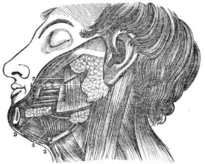
Fig. 58. A view of the salivary glands in their proper situations. 1, The parotid gland. 2, Its duct. 3, The submaxillary gland. 4, Its duct. 5, The sublingual gland, brought to view by the removal of a section of the lower jaw.
238. The PAROTID GLAND, the largest, is situated in front of the external ear, and behind the angle of the jaw. A duct (Steno’s) from this gland opens into the mouth, opposite the second molar tooth of the upper jaw.
237. How many glands about the mouth? Give their names. What does fig. 58 represent? 238. Describe the parotid gland.
239. The SUBMAXILLARY GLAND is situated within the lower jaw, anterior to its angle. Its excretory duct (Wharton’s) opens into the mouth by the side of the fræ´num lin´guæ, (bridle of the tongue.)
240. The SUBLINGUAL GLAND is elongated and flattened, and situated beneath the mucous membrane of the floor of the mouth, on each side of the frænum linguæ. It has seven or eight small ducts, which open into the mouth by the side of the bridle of the tongue.
Observation. In the “mumps,” the parotid gland is diseased. The swelling under the tongue called the “frog” is a disease of the sublingual gland.
Fig. 59.
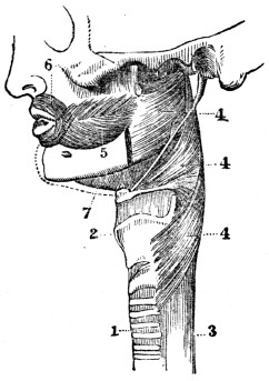
Fig.
59. A side view of the face, œsophagus, and trachea. 1, The trachea (wind pipe.) 2, The larynx. 3, The œsophagus. 4, 4, 4, The muscles of the upper portion of the œsophagus forming the pharynx. 5, The muscle of the cheek. 6, The muscle that surrounds, the mouth. 7, The muscle that forms the floor of the mouth.239. The submaxillary. 240. The sublingual. What observation respecting these glands? What does fig. 59 represent?
241. The PHARYNX is a membranous sac, situated upon the upper portion of the spinal column. It extends from the base of the skull to the top of the tra´che-a, (windpipe,) and is continuous with the œsophagus. From the pharynx are four passages; one opens upward and forward to the nose, the second leads forward to the mouth, the third downward to the trachea and lungs, the fourth downward and backward to the stomach.
242. The ŒSOPHAGUS is a large membranous tube that extends behind the trachea, the heart, and lungs, pierces the diaphragm, and terminates in the stomach. It is composed of two membranes—an internal, or mucous, and a muscular coat. The latter is composed of two sets of fibres; one extends lengthwise, the other is arranged in circular bands.
243. The STOMACH is situated in the left side of the abdomen, immediately below and in contact with the diaphragm. It has two openings; one connected with the œsophagus, called the car´di-ac orifice; the other connected with the upper portion of the small intestine, called the py-lor´ic orifice. It is composed of three coats, or membranes. The exterior or serous coat is very tough and strong, and invests every part of this important organ. The middle, or muscular coat is composed of two layers of muscular fibres, one set of which is arranged longitudinally, the other circularly. The interior coat is called the mucous, and is arranged in ru´gæ, (folds.) The stomach is provided with a multitude of small glands, in which is secreted the gastric fluid.
Illustration. The three coats of the stomach anatomically resemble tripe, which is a preparation of the largest stomach of the cow or ox. The outer coat is smooth and highly polished. The middle coat is composed of minute threads, which are arranged in two layers. The fibres of these layers cross each other. The inner coat is soft, and presents many folds, usually named “the honey-comb.”
241. Describe the pharynx and the passages leading from it. 242. Give the structure of the œsophagus. 243. Where is the stomach situated? How many coats has it? Describe them. What article prepared for food does the stomach resemble?
Fig. 60.
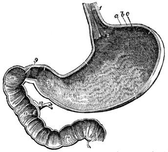
Fig. 60. The inner surface of the stomach and duodenum. 1, The lower portion of the œsophagus. 2, The opening through which the food is passed into the stomach. 8, The stomach. 9, The opening through which the food passes out of the stomach into the duodenum, or upper portion of the small intestine. 10, 11, 14, The duodenum 12, 13, Ducts through which the bile and pancreatic fluid pass into it. a, b, c, The three coats of the stomach.
244. The INTESTINES, or alimentary canal, are divided into two parts—the small and large. The small intestine is about twenty-five feet in length, and is divided into three portions, namely, the Du-o-de´num, the Je-ju´num, and the Il´e-um. The large intestine is about five feet in length, and is divided into three parts, namely, the Cæ´cum, the Co´lon, and the Rec´tum. (Appendix D.)
245. The DUODENUM is somewhat larger than the rest of the small intestine, and has received its name from being in length about the breadth of twelve fingers. It commences at the pylorus, and ascends obliquely backward to the under surface of the liver. It then descends perpendicularly in front of the right kidney, and passes transversely across the lower portion of the spinal column, behind the colon, and terminates in the jejunum. The ducts from the liver and pancreas open into the perpendicular portion, about six inches from the stomach.
244. Explain fig. 60. What is the length of the small intestine, and how is it divided? What is the length of the large intestine? Give its divisions. 245. Describe the duodenum.
246. The JEJUNUM is continuous with the duodenum. It is thicker than the rest of the small intestine, and has a pinkish tinge.
247. The ILEUM is smaller, and thinner in texture, and somewhat paler, than the jejunum. There is no mark to distinguish the termination of the one or the commencement of the other. The ileum terminates near the right haunch-bone, by a valvular opening into the colon at an obtuse angle. This arrangement prevents the passing of substances from the colon into the ileum. The jejunum and ileum are surrounded above and at the sides by the colon.
248. The small intestine, like the stomach, has three coats. The inner, or mucous coat is thrown into folds, or valves. In consequence of this valvular arrangement, the mucous membrane is more extensive than the other tissues, and gives a greater extent of surface with which the aliment comes in contact. There are imbedded under this membrane an immense number of minute glands, and it has a great number of piles, like those upon velvet. For this reason, this membrane is sometimes called the vil´lous coat.
249. The
CÆCUM
is the blind pouch, or cul-de-sac, at the commencement of the large intestine. Attached to its extremity is the ap-pend´ix verm-i-form´is, (a long, worm-shaped tube.) It is from one to six inches in length, and of the size of a goose-quill.What important ducts open into it? 246. Describe the jejunum. 247. The ileum. 248. What is said of the coats of the intestines? Why is the mucous membrane sometimes called the villous coat? 249. Describe the
cæcum
.250. The COLON is divided into three parts—the ascending, transverse, and descending. The ascending colon passes upward from the right haunch-bone to the under surface of the liver. It then bends inward, and crosses the upper part of the abdomen, below the liver and stomach, to the left side under the name of the transverse colon. At the left side, it turns, and descends to the left haunch-bone, and is called the descending colon. Here it makes a peculiar curve upon itself, which is called the sig´moid flex´ure.
Fig. 61.
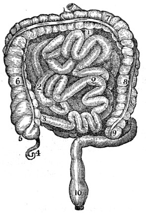
Fig. 61. 1, 1, The duodenum. 2, 2, The small intestine. 3, The junction of the small intestine with the colon. 4, The appendix vermiformis. 5, The
cæcum
. 6, The ascending colon. 7, The transverse colon. 8, The descending colon. 9, The sigmoid flexure of the colon. 10, The rectum.250. Describe the course of the divisions of the colon. Explain fig. 61.
251. The RECTUM is the termination of the large intestine. The large intestine has three coats, like the stomach and small intestine. The longitudinal fibres of the muscular coat are collected into three bands. These bands are nearly one half shorter than the intestine, and give it a sacculated appearance, which is characteristic of the
cæcum
and colon.252. The LACTEALS are minute vessels, which commence in the villi, upon the mucous surface of the small intestine. From the intestine they pass between the membranes of the mes´en-ter-y to small glands, which they enter. The first range of glands collects many small vessels, and transmits a few larger branches to a second range of glands; and, finally, after passing through several successive ranges of these glandular bodies, the lacteals, diminished in number and increased in size, proceed to the enlarged portion of the thoracic duct, into which they open. They are most numerous in the upper portion of the small intestine.
253. The THORACIC DUCT commences in the abdomen, by a considerable dilatation, which is situated in front of the lower portion of the spinal column. From this point, it passes through the diaphragm, and ascends to the lower part of the neck. In its ascent, it lies anterior to the spine, and by the side of the aorta and œsophagus. At the lower part of the neck, it makes a sudden turn downward and forward, and terminates by opening into a large vein which passes to the heart. The thoracic duct is equal in diameter to a goose-quill, and, at its termination, is provided with a pair of semilunar valves, which prevent the admission of venous blood into its cylinder.
251. What is said of the arrangement of the fibres of the muscular coat of the large intestine? 252. What are the lacteals? Give their course from the mucous coat of the intestine to the thoracic duct. 253. Describe the course of the thoracic duct. How is the venous blood prevented from passing into this duct?
[7]
See Chapter XII.
Fig. 62.
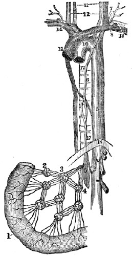
Fig. 62. A portion of the small intestine, lacteal vessels, mesenteric glands, and thoracic duct. 1, The intestine. 2, 3, 4, Mesenteric glands, through which the lacteals pass to the thoracic duct. 5, 6, The thoracic duct. 7, The point in the neck where it turns down to enter the vein at 8. 9, 10, The aorta. 11, 12, Vessels of the neck. 13, 14, 15, The large veins that convey the blood and chyle to the heart. 16, 17, The spinal column. 18, The diaphragm, (midriff.)
Explain fig. 62. What is said respecting the mesenteric glands?
Observation. The mesenteric glands, which are situated between two layers of serous membrane (mesentery) that connects the small intestine with the spinal column, occasionally become diseased in childhood, and prevent the chyle from passing to the thoracic duct. Children thus affected have a voracious appetite, and at the same time are becoming more and more emaciated. The disease is called mesenteric consumption.
254. The LIVER, a gland appended to the alimentary canal, is the largest organ in the system, and weighs about four pounds. It is situated in the right side, below the diaphragm, and is composed of several lobes. Its upper surface is convex; its under, concave. This organ is retained in its place by several ligaments. It performs the double office of separating impurities from the venous blood, and of secreting a fluid (bile) necessary to chylification. On the under surface of the liver is a membranous sac, called the gall-cyst, which is generally considered as a reservoir for the bile.
Fig. 63.
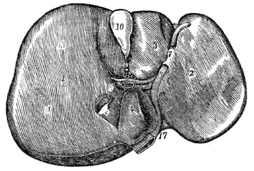
Fig. 63. The under surface of the liver. 1, The right lobe. 2, The left. 3, 4, Smaller lobes. 10, The gall-bladder, or cyst, lodged in its depression. 17, The notch on the posterior border, for the spinal column.
Observation. A good idea of the liver and intestines can be obtained by examining these parts of a pig. In this animal, the sacs, or pouches, of the large intestine are well defined.
255. The PANCREAS is a long, flattened gland, analogous to the salivary glands. It is about six inches in length, weighs three or four ounces, and is situated transversely across the posterior wall of the abdomen, behind the stomach. A duct from this organ opens into the duodenum.
254. Describe the liver. 255. What is said of the pancreas?
256. The SPLEEN, (milt,) so called because the ancients supposed it to be the seat of melancholy, is an oblong, flattened organ, situated in the left side, in contact with the diaphragm, stomach, and the pancreas. It is of a dark, bluish color, and is abundantly supplied with blood, but has no duct which serves as an outlet for any secretion. Its use is not well determined.
Fig. 64.
Fig. 64. The pancreas with its duct, through which the pancreatic secretion passes into the duodenum.
257. The OMENTUM (caul) consists of four layers of the serous membrane, which descends from the stomach and transverse colon. A quantity of adipose matter is deposited around its vessels, which ramify through its structure. Its function is twofold in the animal economy. 1st. It protects the intestines from cold. 2d. It facilitates the movements of the intestines upon each other during their vermicular, or worm-like action.
258. Every part of the digestive apparatus is supplied with arteries, veins, lymphatics, and nervous filaments, from the ganglionic system of nerves.
256. Why is the spleen so called? What is peculiar to this organ? 257. Of what is the omentum composed? What is its use? 258. With what is every part of the digestive apparatus supplied?
CHAPTER XIV.
PHYSIOLOGY OF THE DIGESTIVE ORGANS.
259. Substances received into the stomach as food, must necessarily undergo many changes before they are fitted to form part of the animal body. The solid portions are reduced to a fluid state, and those parts that will nourish the body are separated from the waste material.
260. The first preparation of food for admission into the system, consists in its proper mastication. The lips in front, the cheeks upon the side, the soft palate, by closing down upon the base of the tongue, retain the food in the mouth, while it is subjected to the; process of mas-ti-ca´tion, (chewing.) The tongue rolls the mass around, and keeps it between the teeth, while they divide the food to a fineness suitable for the stomach.
261. While the food is in process of mastication, there is incorporated with it a considerable amount of sa-li´va, (spittle.) This fluid is furnished by the salivary glands, situated in the vicinity of the mouth. The saliva moistens and softens the food, so that, when carried into the pharynx. it is passed, with ease, through the œsophagus into the stomach.
262. When the food has been properly masticated, (and in rapid eaters when it is not,) the soft palate is raised from the base of the tongue backward, so as to close the posterior opening through the nostrils. By a movement of the muscles of the tongue, cheeks, and floor of the mouth, simultaneous with that of the soft palate, the food is pressed into the upper part of the pharynx.
259–272. Give the physiology of the digestive organs. 259. What is necessary before food can nourish the body? 260. Describe how mastication is performed. 261. Of what use is the saliva in the process of mastication? 262. How is the food pressed into the pharynx?
263. When in the pharynx, the food and drink are prevented from passing into the trachea by a simple valve-like arrangement, called the ep-i-glot´tis. The ordinary position of this little organ is perpendicular, so as not to obstruct the passage of air into the lungs; but in the act of swallowing, it is brought directly over the opening of the trachea, called the glot´tis. The food, being forced backward, passes rapidly over the epiglottis into the œsophagus, where the circular band of muscular fibres above, contracts and forces the food to the next lower band. Each band relaxes and contracts successively, and thus presses the alimentary ball downward and onward to the stomach.[8]
Observation. If air is inhaled when the food or drink is passing over the glottis, some portions of it may be carried into the larynx or trachea. This produces violent spasmodic coughing, and most generally occurs when an attempt is made to speak while masticating food; therefore, never talk when the mouth contains food.
263. When the food is in the pharynx, how is it prevented from passing into the trachea, or windpipe? Describe how it is passed into the stomach? Give the observation. 264. Describe how the food in the stomach is converted into chyme.
264. When the food reaches the stomach, the gastric glands are excited to action, and they secrete a powerful solvent, called gastric juice. The presence of food in the stomach also increases a contractile action of the muscular coat, by which the position of the food is changed from one part of this cavity to another. Thus the aliment is brought in contact with the mucous membrane, and each portion of it becomes saturated with gastric juice, by which it is softened, or dissolved into a pulpy homogeneous mass, of a creamy consistence, called Chyme. The food is not all converted into chyme at the same time; but as fast as it is changed, it passes through the pyloric orifice into the duodenum.
Observation. The gastric juice has the property of coagulating liquid albuminous matter when mixed with it. It is this property of rennet, which is an infusion of the fourth stomach of the calf, by which milk is coagulated, or formed into “curd.”
265. The CHYME is conveyed through the pyloric orifice of the stomach into the duodenum. The chyme not only excites an action in the duodenum, but also in the liver and pancreas. Mucus is then secreted by the duodenum, bile by the liver, and pancreatic fluid by the pancreas. The bile and pancreatic fluid are conveyed into the duodenum, and mixed with the chyme. By the action of these different fluids, the chyme is converted into a fluid of a whitish color, called Chyle, and into residuum.
Observation. The bile has no agency in the change through which the food passes in the stomach. In a healthy condition of this organ, no bile is found in it. The common belief, that the stomach has a redundancy of this secretion, is erroneous. If bile is ejected in vomiting, it merely shows, not only that the action of the stomach is inverted, but also that of the duodenum. A powerful emetic will, in this way, generally bring this fluid from the most healthy stomach. A knowledge of this fact might save many a stomach from the evils of emetics, administered on false impressions of their necessity, and continued from the corroboration of these by the appearance of bile, till derangement, and perhaps permanent disease, are the consequences.
266. The CHYLE and residual matter are moved over the mucous surface of the small intestine, by the action of its muscular coat. As the chyle is carried along the tract of the intestine, it comes in contact with the villi, where the lacteal vessels commence. These imbibe, or take up, the chyle, and transfer it through the mesenteric glands into the thoracic duct, through which it is conveyed into a large vein at the lower part of the neck. In this vein the chyle is mixed with the venous fluid. The residual matter is conveyed into the large intestine, through which it is carried and excreted from the system. (Appendix E.)
What peculiar property has gastric juice? 265. Where and how is chyme converted into chyle? What is said in regard to the bile? 266. What becomes of the chyle?
Of the residuum?
267. In the process of digestion, the food is subjected to five different changes. 1st. The chewing and admixture of the saliva with the food; this process is called mastication.
268. 2d. The change through which the food passes in the stomach by its muscular contraction, and the secretion from the gastric glands; this is called chymification.
269. 3d. The conversion of the homogeneous chyme, by the agency of the bile and pancreatic secretions, into a fluid of milk-like appearance; this is chylification.
270. 4th. The absorption of the chyle by the lacteals, and its transfer through them and the thoracic duct, into the subclavian vein at the lower part of the neck.[9]
271. 5th. The separation and excretion of the residuum.
272. Perfection of the second process of digestion requires thorough and slow mastication. The formation of proper chyle demands appropriate mastication and chymification; while a healthy action of the lacteals requires that all the anterior stages of the digestive process be as perfect as possible. (Appendix F.)
267. Recapitulate the five changes in the digestive process.
Note. Let the pupil review the anatomy and physiology of the digestive organs from figs. 62 and 65, or from anatomical outline plate No. 5.
Fig. 65.
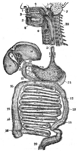
Fig. 65. An ideal view of the organs of digestion, opened nearly the whole
length.
1, The upper jaw. 2, The lower jaw. 3, The tongue. 4, The roof of themouth.
5, The œsophagus. 6, The trachea. 7, The parotid gland. 8, The sublingualgland.
9, The stomach. 10, 10, The liver. 11, The gall-cyst. 12, The duct that conveys the bile to the duodenum, (13, 13.) 14, The pancreas. 15, 15, 15, 15, The small intestine. 16, The opening of the small intestine into the large intestine. 17, 18, 19,20,
The largeintestine.
21, The spleen. 22, The upper part of the spinalcolumn.
CHAPTER XV.
HYGIENE OF THE DIGESTIVE ORGANS.
273. It is a law of the system, that each organ is excited to healthy and efficient action, when influenced by its appropriate stimulus. Accordingly, nutrient food, that is adapted to the wants of the system, imparts a healthy stimulation to the salivary glands during the process of mastication. The food that is well masticated, and has blended with it a
proper
amount of saliva, will induce a healthy action in thestomach.
Well-prepared chyme is the natural stimulus of theduodenum,
liver, and pancreas; pure chyle is the appropriate excitant of the lacteal vessels.274. The perfection of the digestive process, as well as the health of the general system, requires the observance of certain conditions. These will be considered under four heads:—1st. The Quantity of food that should be taken. 2d. Its Quality. 3d. The Manner in which it should be taken. 4th. The Condition of the system when food is taken.
275. The QUANTITY of food necessary for the system varies. Age, occupation, temperament, temperature, habits, amount of clothing, health and disease are among the circumstances which produce the variation.
273–330. Give the hygiene of the digestive organs. 273. Give a law of the system. What is the appropriate stimulus of the salivary glands during mastication? Of the stomach? Of the duodenum? Of the lacteal vessels? 274. What does the perfection of the digestive organs require? 275. What exert an influence on the quantity of food necessary for the system?
276. The child and youth require food to promote the growth of the different parts of the body. The more rapid the growth of the child, the greater the demand for food. This accounts for the keen appetite and vigorous digestion in childhood. When the youth has attained his full growth, this necessity for nutriment ceases; after this period of life, if the same amount of food is taken, and there is no increase of labor or exertion, the digestive apparatus will become diseased, and the vigor of the whole system diminished.
Observation. When the body has become emaciated from want of nutriment, either from famine or disease, there is an increased demand for food. This may be gratified with impunity until the individual has regained the usual size, but repletion should be avoided.
277. Food is required to repair the waste, or loss of substance that attends action. In every department of nature, waste, or loss of substance, attends and follows action. When an individual increases his exercise,—changes from light to severe labor,—or the inactive and sedentary undertake journeys for pleasure, the fluids of the system circulate with increased energy. The old and exhausted particles of matter are more rapidly removed through the action of the vessels of the skin, lungs, kidneys, and other organs, and their places are filled with new atoms, deposited by the small blood-vessels.
278. As the chyle supplies the blood with the newly vitalized particles of matter, there is, consequently, an increased demand for food. This want of the system induces, in general, a sensation of hunger or appetite, which may be regarded as an indication of the general state of the body. The sympathy that exists throughout the system accords to the stomach the power of making known this state to the nervous system, and, if the functions of this faithful monitor have not been impaired by disease, abuse, or habit, the call is imperious, and should be regarded.
276. At what age is the appetite keen and the digestion vigorous? Why? What is said in regard to the quantity of food when the youth has attained his growth? What exception, as given in the observation? 277. Give another demand for food. What effect has increased exercise upon the system?
278.
How are the new particles of matter supplied? What does this induce?279. When exercise or labor is lessened, the quantity of food should be diminished. When a person who has been accustomed to active exercise, or even hard manual labor, suddenly changes to an employment that demands less activity, the waste attendant on action will be diminished in a corresponding degree; hence the quantity of food should be lessened in nearly the same proportion as the amount of exercise is diminished. If this principle be disregarded, the tone of the digestive organs will be impaired, and the health of the system enfeebled.
280. This remark is applicable to those students who have left laborious employments to attend school. Although the health is firm, and the appetite keen from habit, yet every pupil should practise some self-denial, and not eat as much as the appetite craves, the first week of the session. After some days, the real wants of the system will generally be manifested by a corresponding sensation of hunger.
Observation. It is a common observation that in academies and colleges, the older students from the country, who have been accustomed to hard manual labor, suffer more frequently from defective digestion and impaired health than the younger and feebler students from the larger towns and cities.
281. Food is essential in maintaining a proper temperature of the system. The heat of the system, at least in part, is produced in the minute vessels of the several organs, by the union of oxygen with carbon and hydrogen, which the food and drink contain. The amount of heat generated, is greatest when it is most rapidly removed from the system, which occurs in cold weather. This is the cause of the system requiring more food in winter than summer.
279. Why should the quantity of food be diminished when the exercise is lessened? What effect if this principle be disregarded? 280. To what class is this remark applicable? What is often observed among students in academies and colleges? 281. State another demand for food. What is one source of heat in the body?
Observation. Persons that do not have food sufficient for the natural wants of the system, require more clothing than those who are well fed.
282. The last-mentioned principle plainly indicates the propriety and necessity of lessening the quantity of food as the warm season approaches. Were this practised, the tone of the stomach and the vigor of the system would continue unimpaired, the “season complaints” would be avoided, and the “strengthening bitters” would not be sought to create an appetite.
Observation. Stable-keepers and herdsmen are aware of the fact, that as the warm season commences, then animals require less food. Instinct teaches these animals more truly, in this particular, than man allows reason to guide him.
283. The quantity of food should have reference to the present condition of the digestive organs. If they are weakened or diseased, so that but a small quantity of food can be properly digested or changed, that amount only should be taken. Food does not invigorate the system, except it is changed, as has been described in previous paragraphs.
Observation. When taking care of a sick child, the anxiety of the mother and the sufferings of the child may induce her to give food when it would be highly injurious. The attending physician is the only proper person to direct what quantity should be given.
Why do we eat more in the winter than in the summer? What practical observation is given? 282. Why should the quantity of food be lessened as warm weather commences? What would be avoided if this principle were obeyed? 283. Why should the present condition of the digestive organs be regarded in reference to the quantity of food? Mention an instance in which it would be injudicious to give food.
284. The quantity of food is modified, in some degree, by habit. A healthy person, whose exercise is in pure air, may be accustomed to take more food than is necessary. The useless excess is removed from the system by the waste outlets, as the skin, lungs, liver, kidneys, &c. In such cases, if food is not taken in the usual quantity, there will be a feeling of emptiness, if not of hunger, from the want of the usual distention of the stomach. This condition of the digestive organs may be the result of disease, but it is more frequently produced by inordinate daily indulgence in eating, amounting almost to gluttony.
285. Large quantities of food oppress the stomach, and cause general languor of the whole body. This is produced by the extra demands made on the system for an increased supply of blood and nervous fluid to enable the stomach to free itself of its burden. Thus, when we intend to make any extraordinary effort, mental or physical, at least for one meal, we should eat less food than usual, rather than a greater quantity.
286. No more food should be eaten than is barely sufficient to satisfy the appetite. Nor should appetite be confounded with taste. The one is a natural desire for food to supply the wants of the system; the other is an artificial desire merely to gratify the palate.
287. Although many things may aid us in determining the quantity of food proper for an individual, yet there is no certain guide in all cases. It is maintained by some, that the sensation of hunger or appetite is always an indication of the want of food, while the absence of this peculiar sensation is regarded as conclusive evidence that aliment is not demanded. This assertion is not correct, as an appetite may be created for food by condiments and gormandizing, which is as artificial and as morbid as that which craves tobacco or ardent spirits. On the other hand, a structural or functional disease of the brain may prevent that organ from taking cognizance of the sensations of the stomach, when the system actually requires nourishment. Observation shows, that disease, habit, the state of the mind, and other circumstances, exert an influence on the appetite.
284. Show the effect of habit upon the quantity of food that is eaten. What is said in regard to inordinate eating? 285. What is the effect of eating large quantities of food? What suggestion when an extraordinary effort, either mental or physical, is to be made? 286. How much food should generally be eaten? 287. What is the assertion of some persons relative to the quantity of food necessary for the system?
Observation. Dr. Beaumont noticed, in the experiments upon Alexis St. Martin, that after a certain amount of food was converted into chyme, the gastric juice ceased to ooze from the coats of the stomach. Consequently, it has been inferred by some writers on physiology, that the glands which supply the gastric fluid, by a species of instinctive intelligence, would only secrete enough fluid to convert into chyme the aliment needed to supply the real wants of the system. What are the reasons for this inference? There is no evidence that the gastric glands possess instinctive intelligence, and can there be a reason adduced, why they may not be stimulated to extra functional action as well as other organs, and why they may not also be influenced by habit?
288. While all agree that the remote or predisposing cause of hunger is, usually, a demand of the system for nutrient material, the proximate or immediate cause of the sensation of hunger is not clearly understood. Some physiologists suppose that it is produced by an engorged condition of the glands of the stomach which supply the gastric juice; while others maintain that it depends on a peculiar condition of the nervous system.
289. The QUALITY of the food best adapted to the wants of the system is modified by many circumstances. There are many varieties of food, and these are much modified by the different methods of preparation. The same kind of food is not equally well adapted to different individuals, or to the same individual in all conditions; as vocation, health, exposure, habits of life, season, climate, &c., influence the condition of the system.
What does observation show? 288. What is said of the causes of hunger? 289. Why is not the same kind of food adapted to different individuals?
290. All articles of food may be considered in two relations: 1st, As nutritive. 2d, As digestible. Substances are nutritious in proportion to their capacity to yield the elements of chyle, of which carbon, oxygen, hydrogen, and nitrogen are the most essential; they are digestible in proportion to the facility with which they are acted upon by the gastric juice. These properties should not be confounded in the various articles used for food.
291. As a “living body has no power of forming elements, or of converting one elementary substance into another, it therefore follows that the elements of which the body of an animal is composed must be in the food.” (Chap. III.) Of the essential constituents of the human body, carbon, hydrogen, oxygen, and nitrogen are the most important, because they compose the principal part of the animal body; while the other elements are found in very small proportions, and many of them only in a few organs of the system. (Appendix G.)
Observation. Nitrogen renders food more stimulating, particularly if combined with a large quantity of carbon, as beef. Those articles that contain the greatest amount of the constituent elements of the system are most nutritious. As milk and eggs contain all the essential elements of the human system, so they are adapted to almost universal use, and are highly nutritious.
290. In what proportion are substances nutritious? Digestible? Why does beef stimulate the system? What is said of milk and eggs?
292. The following table, by Pereira, in his treatise on Food and Diet may aid the student in approximating to correct conclusions of the quantity of nutriment in different kinds of food, and its adaptation to the wants of the system.
TABLE,
SHOWING THE AVERAGE QUANTITY OF DRY, OR SOLID MATTER, CARBON, NITROGEN, AND MOISTURE, IN DIFFERENT ARTICLES OF DIET.
One hundred Parts.
Dry Matter.
Carbon.
Nitrogen.
Water
Arrowroot,
81.8
36.4
18.2
Beans,
85.89
38.24
14.11
Beef, fresh,
25
12.957
3.752
75
Bread, rye,
67.79
30.674
32.21
Butter,
100
65.6
Cabbage,
7.7
0.28
92.3
Carrot,
12.4
0.30
87.6
Cherries,
25.15
74.85
Chickens,
22.7
77.3
Codfish,
20
80
Cucumbers,
2.86
97.14
Eggs, whites,
20
80
Eggs, yolk,
46.23
53.77
Lard, hog’s,
100
79.098
Milk, cow’s,
12.98
87.02
Oats,
79.2
40.154
1.742
20.8
Oatmeal,
93.4
6.6
Olive-oil,
100
77.50
Oysters,
12.6
87.4
Peaches,
19.76
80.24
Pears,
16.12
83.88
Peas,
84
35.743
16
Plums, greengage,
28.90
71.10
Potatoes,
24.1
10.604
0.3615
75.9
Rye,
83.4
38.530
1.417
16.6
Suet, mutton,
100
78.996
Starch, potato,
82
36.44
18
Starch, wheat,
85.2
37.5
14.8
Sugar, maple,
42.1
Sugar, refined,
42.5
Sugar, brown,
40.88
Turnips,
7.5
3.2175
0.1275
92.5
Veal, roasted,
52.52
14.70
Wheat,
85.5
39.415
1.966
14.5
Note. Let the pupil mention those articles of food that are most nutritious, from a review of this table, and the last four paragraphs.
293. Those articles that do not contain the essential elements of the system should not be used as exclusive articles of diet. This principle has been, and may be illustrated by experiment. Feed a dog with pure sugar, or olive-oil, (articles that contain no nitrogen,) for several weeks, and the evil effects of non-nitrogenous nutriment will be manifested. At first, the dog will take his food with avidity, and seem to thrive upon it; soon this desire for food will diminish, his body emaciate, his eye become ulcerated, and in a few weeks he will die; but mix bran or sawdust with the sugar or oil, and the health and vigor of the animal will be maintained for months. A similar phenomenon will be manifested, if grain only be given to a horse, without hay, straw, or material of like character. (Appendix H.)
294. Some articles of food contain the elements of chyle in great abundance, yet afford but little nutriment, because they are difficult of digestion; while other articles contain but a small quantity of these elements, and afford more nourishment, because they are more easily affected by the digestive process.
293. How has the effect of non-nitrogenous nutriment been illustrated? 294. Why do some articles of food that contain the elements of chyle afford but little nutriment? Why do articles that contain a small quantity of these elements afford more nourishment? 295. How was the time required for digesting different articles of food ascertained?
295. The following table exhibits the general results of experiments made on Alexis St. Martin, by Dr. Beaumont, when he endeavored to ascertain the time required for the digestion of different articles of food.[10] The stomach of St. Martin was ruptured by the bursting of a gun. When he recovered from the effects of the accident under the surgical care of Dr. Beaumont, the stomach became adherent to the side, with an external aperture. Nature had formed a kind of valve, which closed the aperture from the interior, and thus prevented the contents of the stomach from escaping; but on pushing it aside, the process of digestion could be seen. Through this opening, the appearance of the coats of the stomach and food, at different stages of digestion, were examined.
TABLE,
SHOWING THE MEAN TIME OF DIGESTION OF THE DIFFERENT ARTICLES OF DIET.
Articles.
Preparation.
Time
h. m.
Apples, sour, hard,
Raw,
2 50
Apples, sour, mellow,
Raw,
2
Apples, sweet, do.,
Raw,
1 30
Bass, striped, fresh,
Broiled,
3
Beans, pod,
Boiled,
2 30
Beef, fresh, lean, rare,
Roasted,
3
Beef, fresh, lean, dry,
Roasted,
3 30
Beef steak,
Broiled,
3
Beef, with salt only,
Boiled,
3 36
Beef, with mustard,
Boiled,
3 10
Beef, fresh, lean,
Fried,
4
Beef, old, hard, salted,
Boiled,
4 15
Beets,
Boiled,
3 45
Bread, wheat, fresh,
Baked,
3 30
Bread, corn,
Baked,
3 15
Butter,
Melted,
3 30
Cabbage head,
Raw,
2 30
Cabbage, with vinegar,
Raw,
2
Cabbage,
Boiled,
4 30
Cake, sponge,
Baked,
2 30
Carrot, orange,
Boiled,
3 15
Catfish,
Fried,
3 30
Cheese, old, strong,
Raw,
3 30
Chicken, full-grown,
Fricas’d,
2 45
Codfish, cured, dry,
Boiled,
2
Corn, green, & beans,
Boiled,
3 45
Corn bread,
Baked,
3 15
Corn cake,
Baked,
3
Custard,
Baked,
2 45
Dumpling, apple,
Boiled,
3
Ducks, domesticated,
Roasted,
4
Ducks, wild,
Roasted,
4 30
Eggs, fresh,
Boiled hard,
3 30
Eggs, fresh,
Boiled soft,
3
Eggs, fresh,
Fried,
3 30
Eggs, fresh,
Raw,
2
Flounder, fresh,
Fried,
3 30
Fowl, domestic,
Boiled,
4
Fowl, domestic,
Roasted,
4
Goose,
Roasted,
2 30
Lamb, fresh,
Broiled,
2 30
Liver, beef’s, fresh,
Broiled,
2
Meat hashed with vegetables,
Warm’d,
2 30
Milk,
Boiled,
2
Milk,
Raw,
2 15
Mutton, fresh,
Roasted,
3 15
Mutton, fresh,
Broiled,
3
Mutton, fresh,
Boiled,
3
Oysters, fresh,
Raw,
2 55
Oysters, fresh,
Roasted,
3 15
Oysters, fresh,
Stewed,
3 30
Parsnips,
Boiled,
2 30
Pig, sucking,
Roasted,
2 30
Pigs’ feet, soused,
Boiled,
1
Pork, fat and lean,
Roasted,
5 15
Pork, recently salted,
Boiled,
4 30
Pork, recently salted,
Fried,
4 15
Pork, recently salted,
Broiled,
3 15
Pork, recently salted,
Raw,
3
Pork, steak,
Broiled,
3 15
Potatoes, Irish,
Boiled,
3 30
Potatoes, Irish,
Baked,
2 30
Rice,
Boiled,
1
Sago,
Boiled,
1 45
Salmon, salted,
Boiled,
4
Sausage, fresh,
Broiled,
3 20
Soup, beef, vegetables, and bread,
Boiled,
4
Soup, chicken,
Boiled,
3
Soup, mutton,
Boiled,
3 30
Soup, oyster,
Boiled,
3 30
Suet, beef, fresh,
Boiled,
5 30
Suet, mutton,
Boiled,
4 30
Tapioca,
Boiled,
2
Tripe, soused,
Boiled,
1
Trout, salmon, fresh,
Boiled,
1 30
Trout, salmon, fresh,
Fried,
1 30
Turkey, domesticated,
Roasted,
2 30
Turkey,
Boiled,
2 25
Turkey, wild,
Roasted,
2 18
Turnips, flat,
Boiled,
3 30
Veal, fresh,
Broiled,
4
Veal, fresh,
Fried,
4 30
Venison steak,
Broiled,
1 35
296. In view of this table, the question may be suggested, Is that article of food most appropriate to the system which is most easily and speedily digested? To this it may be replied, that the stomach is subject to the same law as the muscles and other organs; exercise, within certain limits, strengthens it. If, therefore, we always eat those articles most easily digested, the digestive powers will be weakened; if over-worked, they will be exhausted. Hence the kind and amount of food should be adapted to the maintenance of the digestive powers, and to their gradual invigoration when debilitated.
Observation. Food that is most easily digested is not always most appropriate to a person convalescing from disease. If the substance passes rapidly through the digestive process, it may induce a recurrence of the disease. Thus the simple preparations which are not stimulating, as water-gruel, are better for a sick person than the more digestible beef and fish.
297. The question is not well settled, whether animal or vegetable food is best adapted to nourish man. There are nations, particularly in the torrid zone, that subsist, exclusively, on vegetables; while those of the frigid zone feed on fish or animal food. In the temperate zone, among civilized nations, a mixed diet is almost universal. When we consider the organization of the human system, the form and arrangement of the teeth, the structure of the stomach and intestines, we are led to conclude, that both animal and vegetable food is requisite, and that a mixed diet is most conducive to strength, health, and long life.
296. How is the question answered, whether that article is most appropriate to the system which is most easily digested? Give observation. 297. What is said of the adaptation of animal and vegetable food to man?
298. The food should be adapted to the distensible character of the stomach and alimentary canal. The former will be full, if it contain only a gill; it may be so distended as to contain a quart. The same is true of the intestines. If the food is concentrated, or contains the quantity of nutriment which the system requires, in small bulk, the stomach and intestines will need the stimulation of distention and friction, which is consequent upon the introduction and transit of the innutritious material into and through the alimentary canal. If the food is deficient in innutritious matter, the tendency is, to produce an inactive and diseased condition of the digestive organs. For this reason, nutrient food should have blended with it innutritious material. Unbolted wheat bread is more healthy than hot flour cakes; ripe fruits and vegetables than rich pies, or jellies.
Observation. 1st. The observance of this rule is of more importance to students, sedentary mechanics, and those individuals whose digestive apparatus has been enfeebled, than to those of active habits and firm health.
2d. The circumstance that different articles of food contain different proportions of waste, or innutritious matter, may be made practically subservient in the following way: If, at any particular season of the year, there is a tendency to a diarrhœa, an article that contains a small proportion of waste should be selected for food; but, if there is a tendency to an inactive or costive condition of the intestinal canal, such kinds of food should be used as contain the greatest proportion of waste, as such articles are most stimulating to the digestive organs, and, consequently, most laxative.
299. In the selection of food, the influence of season and climate should be considered. Food of a highly stimulating character may be used almost with impunity during the cold weather of a cold climate; but in the warm season, and in a warm climate, it would be very deleterious. Animal food, being more stimulating than vegetable, can be eaten in the winter but vegetable food should be used more freely in the spring and summer.
298. What is said of the distensible character of the stomach and alimentary canal? What is the effect of eating highly concentrated food? Why is the unbolted wheat bread more healthy than flour cakes? Give observation 1st. Observation 2d. 299. What kind of food is adapted to cold weather? To warm weather?
300. The influence of food on the system is modified by the age of the individual. The organs of a child are more sensitive and excitable than those of a person advanced in years. Therefore a vegetable diet would be most appropriate for a child, while stimulating animal food might be conducive to the health of a person advanced in life.
Observation. When the digestive organs are highly impressible or diseased, it is very important to adopt a nutritious, unstimulating, vegetable diet, as soon as the warm season commences.
301. Habit is another strong modifying influence. If a person has been accustomed to an animal or vegetable diet, and there is a sudden change from one to the other, a diseased condition of the system, particularly of the digestive apparatus, usually follows. When it is necessary to change our manner of living, it should be done gradually.[11]
302. Some temperaments require more stimulating food than others. As a general rule, those persons whose sensations are comparatively obtuse, and movements slow, will be benefited by animal food; while those individuals whose constitutions are highly impressible, and whose movements are quick and hurried, require a nutritious and unstimulating vegetable diet.
300. What kinds of food are appropriate to old age? Why? What kinds to childhood? Why? 301. What is the effect when there is a sudden change from a vegetable to an animal diet? How should all changes of the system be made? 302. Do different temperaments require different kinds of food? What general rule is given?
[8]
The process of deglutition may be comprehended by analyzing the operation of swallowing food or saliva.
[9]
The chyle is changed by the lacteals and mesenteric glands, but the nature of this change is not, as yet, well defined or understood.
[10]
The time required for the digestion of the different articles of food might vary in other persons; and would probably vary in the same individual at different periods, as the employment, health, season, &c., exert a modifying influence.
[11]
The system is gradually developed, and all changes of food, apparel, labor, exercise, or position, should be gradual. Even a change from a bad to a good habit, on this principle, should be gradual.
CHAPTER XVI.
HYGIENE OF THE DIGESTIVE ORGANS, CONTINUED.
303. The MANNER in which food should be taken is of much practical importance; upon it the health of the digestive organs measurably depends. But few circumstances modify the proper manner of taking food, or should exercise any controlling influence.
304. Food should be taken at regular periods. The interval between meals should be regulated by the character of the food, the age, health, exercise, and habits of the individual. The digestive process is more energetic and rapid in the young, active, and vigorous, than in the aged, indolent, and feeble; consequently, food should be taken more frequently by the former than by the latter class.
305. In some young and vigorous persons, food may be digested in one hour; in other persons, it may require four hours or more. The average time, however, to digest an ordinary meal, will be from two to four hours. In all instances, the stomach will require from one to three hours to recruit its exhausted powers after the labor of digesting a meal before it will again enter upon the vigorous performance of its duties.
306. Food should not be taken too frequently. If food is taken before the stomach has regained its tone and energy by repose, the secretion of the gastric juice, and the contraction of the muscular fibres, will be imperfect. Again, if food is taken before the digestion of the preceding meal has been completed, the effects will be still worse, because the food partially digested becomes mixed with that last taken.
Therefore
the interval between each meal should be long enough for the whole quantity to be digested, and the time of repose should be sufficient to recruit the exhausted organs. The feebler the person and the more debilitated the stomach, the more important to observe the above directions.303. Why is it important that we regard the manner of taking our food? 304. How should the intervals between meals be regulated? 305. What is the average time required to digest an ordinary meal? 306. Why should not food be taken too frequently?
Observation. In the feeding of infants, as well as in supplying food to older children, the preceding suggestions should always be regarded. The person who has been confined by an exhausting sickness, should most scrupulously regard this rule, if he wishes to regain his strength and flesh with rapidity. As the rapidity of the digestive process is less in students and individuals who are engaged in sedentary employments, than in stirring agriculturists, the former class are more liable to take food too frequently than the latter, while its observance is of greater importance to the sedentary artisan than to the lively lad and active farmer.
307. Food should be well masticated. All solid aliments should be reduced to a state of comparative fineness, by the teeth, before it is swallowed; the gastric fluid of the stomach will then blend with it more readily, and act more vigorously in reducing it to chyme. The practice of swallowing solid food, slightly masticated, or “bolting” it down, tends to derange the digestive process and impair the nutrition of the system.
308. Mastication should be moderate, not rapid. In masticating food, the salivary glands are excited to action, and some time must elapse before they can, secrete saliva in sufficient quantities to moisten it. If the aliment is not supplied with saliva, digestion is retarded; besides, in rapid eating, more food is generally consumed than the system demands, or can be easily digested. Laborers, as well as men of leisure, should have ample time for taking their meals. Imperfect mastication is a prevailing cause of indigestion.
What persons would be benefited by observing the preceding remarks? 307. Why should food be well masticated? What is the effect of “bolting down” food? 308. How should mastication be performed? Why?
309. Food should be masticated and swallowed without drink. As the salivary glands supply fluid to moisten the dry food, the use of tea, coffee, water, or any other fluid, is not demanded by nature’s laws while taking a meal. One objection to “washing down” the food with drink is, the aliment is moistened, not with the saliva, but with the drink. This tends to induce disease, not only in the salivary organs, by leaving them in a state of comparative inactivity, but in the stomach, by the deficiency of the salivary stimulus. Another is, large quantities of fluids, used as drinks, give undue distention to the stomach, and lessen the energy of the gastric juice by its dilution, thus retarding digestion. Again, drinks taken into the stomach must be removed by absorption before the digestion of other articles is commenced.
Observation. Were it customary not to place drinks on the table until the solid food is eaten, the evil arising from drinking too much at meals would be obviated. The horse is never known to leave his provender, nor the ox his blade of grass, to wash it down; but many persons, from habit rather than thirst, drink largely during meals.
310. The peculiar sensation in the mouth and fauces, called thirst, may not always arise from the demand for fluids to increase the serum (water) of the blood, as in the desire for drink attendant on free perspiration, for then, pure water or some diluent drink is absolutely necessary; but it may be the result of fever, or local disease of the parts connected with the throat. In many instances, thirst may be allayed by chewing some hard substance, as a dry cracker. This excites a secretion from the salivary glands, which removes the disagreeable sensation. In thirst, attendant on a heated condition of the system, this practice affords relief, and is safe; while the practice of drinking large quantities of cold fluids, is unsafe, and should never be indulged.
Why should all persons have ample time for eating? 309. Why are drinks not necessary while masticating food? Give the objections to “washing down” food. What observation relative to drink? 310. Does the sensation of thirst always arise from a real want of the system?
311. Food or drink should not be taken when very hot. When food or drink is taken hot, the vessels of the mucous membrane of the gums, mouth, and stomach are unduly stimulated for a short time; and this is followed by reaction, attended by a loss of tone, and debility of these parts. This practice is a fruitful cause of spongy gums, decayed teeth, sore mouth, and indigestion.
312. Food or drink should not be taken very cold. If a considerable quantity of very cold food or liquid be taken immediately into the stomach, the health will be endangered, and the tone of the system will be impaired, from the sudden abstraction of heat from the coats of the stomach, and from surrounding organs, to impart warmth to the cold food or drink. This arrests the digestive process, and the food is retained in the stomach too long, and causes oppression and irritation. Consequently, food and drink that are moderately heated are best adapted to the natural condition of the digestive apparatus.
Observation. Food of an injurious quality, or taken in an improper manner, affects the inferior animals as well as man. The teeth of cows that are closely penned in cities, and are fed on distillery slops, or the unhealthy slops and remnants of kitchens, decay and fall out in about two years. Can the milk of such diseased animals be healthy—the proper nourishment for children?
Give instances when it does and when it does not. 311. Why should not food or drink be taken hot? 312. Why should they not be taken cold? Show some of the effects of improper food upon the inferior
animals.
313. The CONDITION of the system should be regarded when food is taken. This is necessary, as the present and ulterior condition of the digestive apparatus is strongly influenced by the state of the other organs of the system.
314. Food should not be taken immediately after severe exertion, either of the body or mind. For all organs in action require and receive more blood and nervous fluid, than when at rest. This is true of the brain, muscles, and vocal organs, when they have been actively exercised. The increased amount of fluid, both sanguineous and nervous, supplied to any organ during extra functional action, is abstracted from other parts of the system. This enfeebles and prostrates the parts that supply the blood and nervous fluid to the active organ. Again, when any organ has been in vigorous action for a few hours, some time will elapse before the increased action of the arteries and nerves abates, and a due supply of fluids is transmitted to other organs, or an equilibrium of action in the system is reëstablished.
315. Thus food should not be taken immediately after severe mental labor, protracted speaking, continued singing, or laborious manual toil; as the digestive organs will be in a state of comparative debility, and consequently unfit to digest food. From thirty to sixty minutes should elapse, after the cessation of severe employment, before food is taken. This time may be spent in cheerful amusement or social conversation.
Observation. The practice of students and accountants going immediately from severe mental labor to their meals, is a pernicious one, and a fruitful cause of indigestion and mental debility. The custom of farmers and mechanics hurrying from their toil to the dinner-table, does much to cause dyspepsia and debility among these classes in community.
313. Should the condition of the system be regarded in taking food? 314. When should food not be taken? Why? What is the result when an organ has been in vigorous action? 315. After the cessation of severe toil, how much time should expire before eating? What is one cause of indigestion among students and accountants?
316. Severe mental or manual toil should not be entered upon immediately after eating. As there is an increased amount of blood and nervous fluid supplied to the stomach and alimentary canal during the digestion of food, a deficiency exists in other organs. This is evinced by a slight paleness of the skin, and a disinclination to active thought and exercise. Under such circumstances, if either the mind, vocal organs, or muscles are called into energetic action, there will be an abstraction of the necessary amount of blood and nervous fluid from the stomach, and the process of digestion will be arrested. This will not only cause disease of the digestive organs, but chyle will not be formed, to nourish the system.
Illustration. An English gentleman fed two dogs upon similar articles of food. He permitted one to remain quiet in a dark room; the other he sent in pursuit of game. At the expiration of one hour, he had both killed. The stomach of the dog that had remained quiet was nearly empty. The food had been properly changed and carried forward into the alimentary canal. In the stomach of the dog that had used his muscles in chasing game, the aliment remained nearly unaltered.
317. The same principle may be applied to the action of the organs of man. If his mind or muscles act intensely soon after eating, the stomach will not be sufficiently stimulated by blood and nervous fluid to change the food in a suitable period. The Spanish practice of having a “siesta,” or sleep after dinner, is far better than the custom of the Anglo-Saxon race, who hurry from their meals to the field, shop, or study, in order to save time, which, in too many instances, is lost by a sense of oppression and suffering which soon follows.
316. Why should not severe manual or mental exertion be made immediately after eating? State the illustration. 317. May this principle be applied to the action of the human stomach? What is said of the Spanish custom of resting after dinner?
318. In some instances of good health, the infringement of this organic law may seem to pass with impunity, but Nature, though lenient, sooner or later asserts her claims. The practice of the Spaniard may be improved by indulging, for an hour before resuming toil, in moderate exercise of the muscular system, conjoined with agreeable conversation and a hearty laugh, as this facilitates digestion, and tends to “shake the cobwebs from the brain.”
Observation. No judicious teamster drives his animals as soon as they have swallowed their food, but gives them a period for repose, so that their food may be digested, and their systems invigorated. In this way, he secures the greatest amount of labor from his team.
319. The mind exerts an influence upon the digestive process. This is clearly exhibited, when an individual receives intelligence of the loss of a friend or of property. He may at the time be sitting before a plentiful board, with a keen appetite; but the unexpected news destroys it, because the excited brain withholds its stimulus. This shows the propriety of avoiding absorbing topics of thought at meals, as labored discussions and matters of business; but substitute cheerful and light conversation, enlivening wit, humor, the social intercourse of family and friends; these keep the brain in action, but not in toil. Under such circumstances, the blood and nervous fluid flow freely, the work of digestion is readily commenced, and easily carried on.
320. Indigestion arising from a prostration of the nervous system, should be treated with great care. The food should be simple, nutritious, moderate in quantity, and taken at regular periods. Large quantities of stimulating food, frequently taken, serve to increase the nervous prostration. Those afflicted should exercise in the open air, and engage in social conversation, that the brain may be excited to a natural or healthy action, in order that it may impart to the digestive organs the necessary stimulation.
Of the Anglo-Saxon race? 318. How can the Spanish custom be improved? 319. How is the influence of the mind on the digestive process exhibited? What does it show the necessity of avoiding? 320. How should indigestion arising from nervous prostration be treated?
321. Persons should abstain from eating, at least three hours before retiring for sleep. It is no unusual occurrence, for those persons who have eaten heartily immediately before retiring to sleep, to have unpleasant dreams, or to be aroused from their unquiet slumber by colic pains. In such instances, the brain becomes partially dormant, and does not impart to the digestive organs the requisite amount of nervous influence. The nervous stimulus being deficient, the unchanged food remains in the stomach, causing irritation of this organ.
Illustration. A healthy farmer, who was in the habit of eating one fourth of a mince pie immediately before going to bed, became annoyed with unpleasant dreams, and, among the varied images of his fancy, he saw that of his deceased father. Becoming alarmed, he consulted a physician, who, after a patient hearing of the case, gravely advised him to eat half of a mince pie, assuring him that he would then see his grandfather.
322. When the general system and digestive organs are enfeebled, mild, unstimulating food, in small quantities, should be given. In the instance of a shipwrecked and famished mariner, or a patient recovering from disease, but a small quantity of nourishment should be given at a time. The reason for this, is, that when the stomach is weakened from want of nourishment, it is as unfitted for a long period of action in digesting food, as the muscles are, under like circumstances, for walking. Consequently, knowledge and prudence should direct the administration of food under these circumstances. The popular adage, that “food never does harm when there is a desire for it,” is untrue, and, if practically adopted, may be injurious and destructive to life.
321. What is the effect of eating immediately before retiring for sleep? How is this illustrated in the case of a healthy farmer? 322. How should the food be given when both the digestive organs and general system are debilitated? Give the reason.
Observation. Liquids are rapidly removed from the stomach by absorption. Hence, in cases of great prostration, when it is desirable to introduce nutriment into the system, without delay, the animal and vegetable broths are a desirable and convenient form of supplying aliment.
323. The condition of the skin exercises an important influence on the digestive apparatus. Let free perspiration be checked, either from uncleanliness or from chills, and it will diminish the functional action of the stomach and its associated organs. This is one of the fruitful causes of the “liver and stomach complaints” among the half-clothed and filthy population of the crowded cities and villages of our country. Attention to clothing and bathing would likewise prevent many of the diseases of the alimentary canal, called “season complaints,” particularly among children.
324. Restricting the movements of the ribs and diaphragm impairs digestion. At each full inspiration, the ribs are elevated, and the central portion of the diaphragm is depressed, from one to two inches. This depression is accompanied by a relaxation of the anterior abdominal walls. At each act of expiration, the relaxed abdominal muscles contract, the ribs are depressed, the diaphragm relaxes, and its central parts ascend. These movements of the midriff cause the elevation and depression of the stomach, liver, and other abdominal organs, which is a natural stimulus of these parts.
In cases of great prostration, what is recommended? 323. How is the influence that the skin exercises on the digestive organs illustrated? 324. What effect on the digestive process has the restriction of the ribs and diaphragm?
325. It is noted of individuals who restrain the free movements of the abdominal muscles by tight dresses, that the tone and vigor of the digestive organs are diminished. The restricted waist will not admit of a full and deep inspiration and so essential is this to health, that abuse in this respect soon enfeebles and destroys the functions of the system.
326. Pure air is necessary to give a keen appetite and vigorous digestion. The digestive organs not only need the stimulus of blood, but they absolutely need the influence of pure blood, which cannot exist in the system, except when we breathe a pure air. From this we learn why those persons who sleep in small, ill ventilated rooms, have little or no appetite in the morning, and why the mouth and throat are so dry and disagreeable. The effect of impure blood, in diminishing the desire for food, and enfeebling the digestive organs, is well illustrated by the following incidents.
Illustrations. 1st. Dr. Reid, in his work on “Ventilation of Rooms,” relates that an innkeeper in London, when he provided a public dinner, always spread his tables in an under-ground room, with low walls, where the air was confined and impure. He assigned as a reason for so doing, that his guests consumed only one third as much food and wine, as if the tables were laid in the open air.
2d. A manufacturer stated before a committee of the British Parliament, that he had removed an arrangement for ventilating his mill, because he noticed that his men ate much more after his mill was ventilated, than previous to admitting fresh air into the rooms, and that he could not afford to have them breathe pure air.
Observation. Many of the cases of indigestion among clergymen, seamstresses, school teachers, sedentary mechanics, and factory operatives, are produced by breathing the impure air of the rooms they occupy. These cases can be prevented, as well as cured, by proper attention to ventilation.
325. What is observed of those individuals that restrict the movements of the abdominal muscles? 326. Why is pure air necessary to vigorous digestion? Give illustration 1st. Illustration 2d. What is one cause of indigestion among the sedentary class in community?
327. The position of a person, in standing or sitting, exerts an influence upon the digestive organs. If a person lean, or stoop forward, the distance between the pelvic bones and the diaphragm is diminished. This prevents the depression of the diaphragm, while the stomach, liver, pancreas, and other abdominal organs, suffer compression, which induces many severe diseases of these organs. As healthy and well-developed muscles keep the spinal column in an erect position, which conduces to the health of the organs of digestion, the child should be taught to avoid all positions but the erect, while studying or walking. This position, combined with unrestricted waists, will do much to remove the now prevalent disease, dyspepsia.
328. Whatever kind of aliment is taken, it is separated into nutriment and residuum; the former of which is conveyed, through the medium of the circulation, to all organs of the system, and the latter, if not expelled, accumulates, causing headache and dizziness, with a general uneasiness; and, if allowed to continue, it lays the foundation of a long period of suffering and disease. For the preservation of health, it is necessary that there should be a daily evacuation of the residual matter.
Observation. In chronic diseases of the digestive organs, very frequently, there is an inactive, or costive condition of the alimentary canal. This may be removed in many cases, and relieved in all instances, by friction over the abdominal organs, and by making an effort at some stated period each day, (evening is best,) to evacuate the residuum. In acute diseases, as fever, regard should be given to regularity in relieving the intestines of residuum. Attention to this suggestion will in many instances obviate the necessity of cathartic medicine.
327.
Why does the position of a person affect digestion? 328. Into what are different kinds of aliment separated?329. We would add, for the benefit of those afflicted with hemorrhoids, or piles, that the best time for evacuating the intestinal canal would be immediately before retiring for the night. During the night, while recumbent, the protruding parts return to their proper place, and the surrounding organs acquire increased tone to retain them. The same observance will do much to prevent such prostrating diseases.[12]
330. To recapitulate: digestion is most perfect when the action of the cutaneous vessels is energetic; the brain and vocal organs moderately stimulated by animated conversation; the blood well purified; the muscular system duly exercised; the food of an appropriate quality, taken in proper quantities, at regular periods, and also properly masticated.
330. Give the summary when digestion is most perfect.
CHAPTER VXII.
THE CIRCULATORY ORGANS.
331. The ultimate object of the food and drink introduced into the body, is to furnish material to promote the growth and repair the waste of the organs of the system. The formation of chyle (the nutrient portion of the food) has been traced through the digestive process, and its transfer into the vein at the lower part of the neck, from which it is conveyed to the heart; and, finally, in the lungs it assimilates to the character of blood.
332. The BLOOD, after standing a short time, when drawn from its vessels, separates into se´rum, (a watery fluid,) and co-ag´u-lum, (clot.) This fluid is distributed to every part of the system. There is no part so minute that it does not receive blood. The organs by which this distribution is effected are so connected that there is properly neither beginning nor end; but as it respects their functions, they are connected in a complete circle. From this circumstance, they are called the Circulatory Organs.
ANATOMY OF THE CIRCULATORY ORGANS.
333. The CIRCULATORY ORGANS are the Heart,
Ar´te-ries,
Veins, and Cap´il-la-ries.334. The HEART is placed obliquely, in the left cavity of the chest, between the right and left lung. Its general form is that of an inverted cone, the base of which is directed upward and backward, toward the right shoulder, while its apex points forward to the left side, about three inches from the sternum to the space between the fifth and sixth ribs. Its under side rests upon the tendinous portion of the diaphragm. The heart is surrounded by a sac, called the per-i-car´di-um, (heart-case.) The interior surface of this membrane secretes a watery fluid, that lubricates the exterior of the heart, and obviates friction between it and the pericardium.
331. what is the ultimate object of the food? 332. Of what is the blood composed? What is said of the distribution of the blood? 333. Name the circulatory organs. 334–351. Give the anatomy of the circulatory organs. 334. Describe the heart.
Fig. 66.
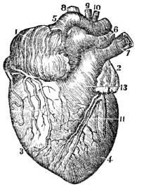
Fig. 67.
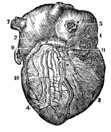
Fig. 66. A front view of the heart. 1, The right auricle of the heart. 2, The left auricle. 3, The right ventricle. 4, The left ventricle. 5, 6, 7, 8, 9, 10, The vessels[13] through which the blood passes to and from the heart.
Fig. 67. A back view of the heart. 1, The right auricle. 2, The left auricle. 3, The right ventricle. 4, The left ventricle. 5, 6, 7, The vessels that carry the blood to and from the heart. 9, 10, 11, The nutrient vessels of the heart.
With what is it surrounded? What is its use? How much fluid does this membrane contain when healthy?
Observation. In health, there is usually about a tea-spoonful of fluid in the pericardium. When these parts are diseased, it may be thrown out more abundantly, and sometimes amounts to several ounces, producing a disease called dropsy of the heart. But all the unpleasant sensations in the region of the heart are not caused by an increased amount of fluid in the pericardium, as this disease is not of frequent occurrence.
335. The heart is composed of muscular fibres, that traverse it in different directions, some longitudinally, but most of them in a spiral direction. The human heart is a double organ, or it has two sides, called the right and the left. The compartments of the two sides are separated by a muscular sep´tum, or partition. Again, each side of the heart is divided into two parts, called the Au´ri-cle (deaf ear) and the Ven´tri-cle.
Fig. 68
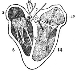
Fig. 68. A section of the heart, showing its cavities and valves. 3, The right auricle. 4, The opening between the right auricle and right ventricle. 5, The right ventricle. 6, The tricuspid valves. 7, The pulmonary artery. 9, The semilunar valves of the pulmonary artery. 10, The septum between the right and left ventricle. 12, The left auricle. 13, The opening between the left auricle and left ventricle. 14, The left ventricle. 15, The mitral valves. 16, The aorta. 17, The semilunar valves of the aorta.
336. The AURICLES differ in muscularity from the ventricles. Their walls are thinner, and of a bluish color. These cavities are a kind of reservoir, designed to contain the blood arriving by the veins.
337. The VENTRICLES not only have their walls thicker than the auricles, but they differ in their internal structure. From the interior of these cavities arise fleshy columns, called co-lum´næ car´ne-æ. The walls of the left ventricle are thicker and stronger than those of the right.
335. Of what is the heart composed? Give its divisions. 336. Describe the auricles. 337. Describe the ventricles.
338. The cavities in the right side of the heart are triangular in shape; those of the left, oval. Each cavity will contain about two ounces of blood. Between the auricle and ventricle in the right side of the heart, there are three folds, or doublings, of thin, triangular membrane, called the tri-cus´pid valves. Between the auricle and ventricle in the left side, there are two valves, called the mi´tral. There are seen passing from the floating edge of these valves to the columnæ carneæ, small white cords, called chor´dæ ten´di-næ, which prevent the floating edge of the valve from being carried into the auricle.
339. The right ventricle of the heart gives rise to the Pul´mo-na-ry artery; the left ventricle, to a large artery called the A-ort´a. At the commencement of each of these arteries there are three folds of membrane, and from their shape, they are called sem-i-lu´nar valves.
340. The heart is supplied with arteries and veins, which ramify between its muscular fibres, through which its nutrient blood passes. It has, likewise, a few lymphatics, and many small nervous filaments from the sympathetic system of nerves. This organ, in its natural state, exhibits but slight indications of sensibility, and although nearly destitute of the sensation of touch, it is yet, however, instantly affected by every painful bodily excitement, or strong mental emotions.
Observation. To obtain a clear idea of the heart and its valves, it is recommended to examine this part of an ox or calf. In order that each ventricle be opened without mutilating the fleshy columns, tendinous cords, and valves, cut on each side of the septum parallel to it. This may be easily found between the ventricles, as they differ in thickness.
338. How do the cavities in the heart differ? What is found between the auricle and ventricle in the right side of the heart? How many valves in the left side, and their names? Where are the tendinous cords, and what is their use? 339. What vessels proceed from the ventricles? What is said of their valves? 340. With what is the heart supplied? What is said of its sensibility? How can an idea of the structure of the heart be obtained?
341. The ARTERIES are the cylindrical tubes that convey the blood from the heart to every part of the system. They are dense in structure, and preserve, for the most part, the cylindrical form, when emptied of their blood, which is their condition after death.
342. The arteries are composed of three coats. The external, or cellular coat, is firm and strong; the middle, or fibrous coat, is composed of yellowish fibres. This coat is elastic, fragile, and thicker than the external coat. Its elasticity enables the vessel to accommodate itself to the quantity of blood it may contain. The internal coat is a thin, serous membrane, which lines the interior of the artery, and gives it the smooth polish which that surface presents. It is continuous with the lining membrane of the heart.
343. Communications between arteries are free and numerous. They increase in frequency with diminution in the size of the branches, so that through the medium of the minute ramifications, the entire body may be considered as one circle of inosculation. The arteries, in their distribution through the body, are enclosed in a loose, cellular investment, called a sheath, which separates them from the surrounding tissues.
344. The PULMONARY ARTERY commences in front of the origin of the aorta. It ascends obliquely to the under surface of the arch of the aorta, where it divides into two branches, one of which passes to the right, the other to the left lung. These divide and subdivide in the structure of the lungs, and terminate in the capillary vessels, which form a net-work around the air-cells, and become continuous with the minute branches of the pulmonary veins. This artery conveys the impure blood to the lungs, and, with its corresponding veins, establishes the lesser, or pulmonic circulation.
341. What are arteries? 342. Give their structure. 343. What is said of the communications between the arteries? In their distribution, how are they separated from the surrounding tissues? 344. Describe the pulmonary artery.
Fig. 69.
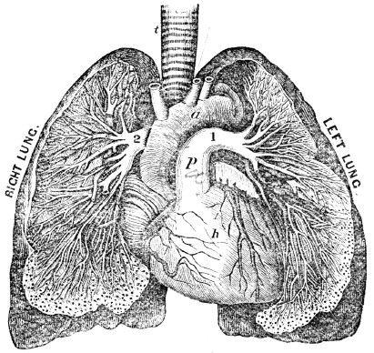
Fig. 69. t, The trachea. h, The heart. a, The aorta. p, The pulmonary artery 1, The branch of the pulmonary artery that divides in the left lung. 2, The branch that divides in the right lung.
The divisions of this artery continue to divide and subdivide, until they become no larger than hairs in size. These minute vessels pass over the air-cells, represented by small dark points around the margin of the lungs.
345. The AORTA proceeds from the left ventricle of the heart, and contains the pure, or nutrient blood. This trunk gives off branches, which divide and subdivide to their ultimate ramifications, constituting the great arterial tree which pervades, by its minute subdivisions, every part of the animal frame. This great artery and its divisions, with their returning veins, constitute the greater, or systemic circulation.
What does this artery and its corresponding veins establish? Explain fig. 69. 345. Describe the aorta. What do this artery and its corresponding veins constitute?
Fig. 70.
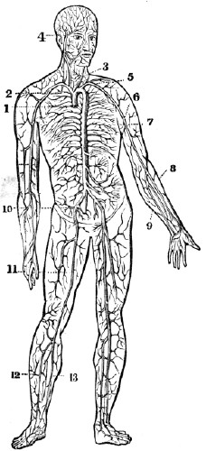
Fig. 70. The aorta and its branches. 1, The commencement of the aorta. 2, The arch of the aorta. 3, The carotid artery. 4, The temporal artery. 5, The subclavian artery. 6, The axillary artery. 7, The brachial artery. 8, The radial artery. 9, The ulnar artery. 10, The iliac artery. 11, The femoral artery. 12, The tibial artery, 13. The peroneal artery.
346. The VEINS are the vessels which return the blood to the auricles of the heart, after it has been circulated by the arteries through the various tissues of the body. They are thinner and more delicate in structure than the arteries, so that when emptied of their blood, they become flattened and collapsed. The veins commence by minute radicles in the capillaries, which are every where distributed through the textures of the body, and coalesce to constitute larger and larger branches, till they terminate in the large trunks which convey the dark-colored blood directly to the heart. In diameter they are much larger than the arteries, and, like those vessels, their combined area would constitute an imaginary cone, the apex of which is placed at the heart, and the base at the surface of the body.
What does fig. 70 represent? 346. What are the veins?
347. The communications between the veins are more frequent than between the arteries, and take place between the larger as well as among the smaller vessels. The office of these inosculations is very apparent, as tending to obviate the obstructions to which the veins are peculiarly liable, from the thinness of their coats, and from inability to overcome great impediments by the force of their current. These tubes, as well as the arteries, are supplied with nutrient vessels, and it is to be presumed that nervous filaments from the sympathetic nerves are distributed to their coats.
348. The external, or cellular coat of the veins, is dense and firm, resembling the cellular tunic of the arteries. The middle coat is fibrous, like that of the arteries, but extremely thin. The internal coat is serous, and also similar to that of the arteries. It is continuous with the lining membrane of the heart at one extremity, and with the lining membrane of the capillaries at the other.
349. At certain intervals, the internal coat forms folds, or duplicatures, which constitute valves. They are generally composed of two semilunar folds, one on each side of the vessel. The free extremity of the valvular folds is concave, and directed forward, so that while the current of blood sets toward the heart, they present no impediment to its free passage; but let the current become retrograde, and it is impeded by their distention. The valves are most numerous in the veins of the extremities, particularly the deeper veins situated between the muscles; but in some of the larger trunks, and also in some of the smaller veins, no valves exist.
Where do they commence? 347. What is said of their communications? What is the apparent design of the inosculations of the veins? What vessels are distributed to the coats of the veins? 348. Give the structure of the coats of the veins. 349. How are the valves in the veins formed?
Fig. 71.
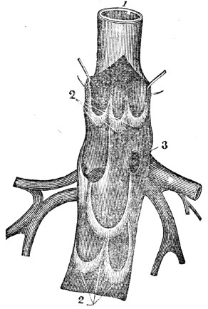
Fig. 71. A vein laid open to show the valves. 1, The trunk of the vein. 2, 2, Its valves. 3, An opening of a branch into the main trunk.
What is their use? Where are they the most numerous?
350. The CAPILLARIES constitute a microscopic net-work, and are so distributed through every part of the body as to render it impossible to introduce the smallest needle beneath the skin, without wounding several of these fine vessels. They are remarkable for the uniformity of diameter, and for the constant divisions and communications which take place between them.
351. The capillaries inosculate, on the one hand, with the terminal extremity of the arteries, and on the other, with the commencement of the veins. They establish the communication between the termination of the arteries and the beginning of the veins. The important operations of secretion and the conversion of the nutrient materials of the blood into bone, muscle, &c., are performed in these vessels.
Fig. 72.
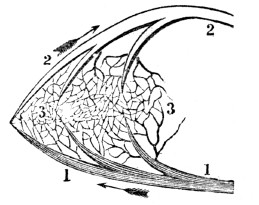
Fig. 73.
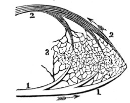
Fig. 72. An ideal view of a portion of the pulmonic circulation. 1, 1, A branch of the artery that carries the impure blood to the lungs. 3, 3, Capillary vessels. 2, 2, A vein through which red blood is returned to the left side of the heart.
Fig. 73. An ideal view of a portion of the systemic circulation. 1, 1, A branch of the aorta. This terminates in the capillaries, (3, 3.) 2, 2, A vein through which the impure blood is carried to the right side of the heart.
350. What do the capillaries constitute? For what are they remarkable? 351. What relation do they bear to the arteries and veins? What important operations are performed in these vessels? What is represented by fig. 72? By fig. 73?
CHAPTER XVIII.
PHYSIOLOGY OF THE CIRCULATORY ORGANS.
352. The walls of all the cavities of the heart are composed of muscular fibres, which are endowed with the property of contracting and relaxing, like the muscles of the extremities. The contraction and relaxation of the muscular tissue of the heart, produce a diminution and enlargement of both auricular and ventricular cavities. The auricles contract and dilate simultaneously, and so do the ventricles; yet the contraction and dilatation of the auricles do not alternate with the contraction and dilatation of the ventricles, as the dilatation of the one is not completed before the contraction of the other commences. The dilatation of the ventricles is termed the di-as´to-le of the heart; their contraction, its sys´to-le.
353. The ventricles contract quicker and more forcibly than the auricles, and they are three times longer in dilating than contracting. The walls of the right ventricle, being thinner than the left, are more distensible, and thus this cavity will contain a greater amount of blood. This arrangement adapts it to the venous system, which is more capacious than the arterial. The thicker and more powerful walls of the left ventricle adapt it to expel the blood to a greater distance.
354. The valves in the heart permit the blood to flow from the auricles to the ventricles, but prevent its reflowing. The valves at the commencement of the aorta and pulmonary artery, permit the blood to flow from the ventricles into these vessels, but prevent its returning.
352–366. Give the physiology of the circulatory organs. 352. What do the contraction and relaxation of the muscular walls of the heart produce? How do the auricles and ventricles contract and dilate? 353. What is said of the contraction and dilatation of the ventricles in the heart? How is the right ventricle adapted to its function? How the left? 354. What is the use of the valves in the heart? Those of the aorta and pulmonary artery?
355. The function of the different parts of the heart will be given, by aid of fig. 74. The blood passes from the right auricle (3) into the right ventricle, (5,) and the tricuspid valves (6) prevent its reflux; from the right ventricle the blood is forced into the pulmonary artery, (7,) through which it passes to the lungs. The semilunar valves (9) prevent this circulating fluid returning to the ventricle. The blood, while passing over the air-cells in the lungs, in the minute divisions of the pulmonary artery, is changed from a bluish color to a bright red. It is then returned to the left auricle of the heart by the pulmonary veins, (11, 11.)
[12]
The urinary organs, as well as the intestinal canal, should be frequently and regularly evacuated. Some most distressing and frequently incurable complaints are caused by false customs and false delicacy in this particular. Teachers should be particularly careful, and regard this suggestion in reference to young pupils.
[13]
All vessels that carry blood to the heart, are called veins. All vessels that carry blood from the heart, are called arteries.
Fig. 74.
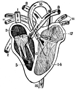
Fig. 74. 1, The descending vena cava, (vein.) 2, The ascending vena cava, (vein.) 3, The right auricle. 4, The opening between the right auricle and the right ventricle. 5, The right ventricle. 6, The tricuspid valves. 7, The pulmonary artery. 8, 8, The branches of the pulmonary artery that pass to the right and left lung. 9, The semilunar valves of the pulmonary artery. 10, The septum between the two ventricles of the heart. 11, 11, The pulmonary veins. 12, The left auricle. 13, The opening between the left auricle and ventricle. 14, The left ventricle. 15, The mitral valves. 16, 16, The aorta. 17, The semilunar valves of the aorta.
355. Describe the course of the blood from the right auricle in the heart to the lungs.
Observation. If the blood is not changed in the lungs, it will not flow to the pulmonary veins. This phenomenon is seen in instances of death from drowning, strangling, carbonic acid, &c. The same is true, but in a less degree, of individuals whose apparel is tight, as well as of those who breathe impure air, or have diseased lungs.
356. The left auricle, (12,) by its contraction, forces the blood into the left ventricle, (14.) The mitral valves (15) prevent its reflowing. From the left ventricle the blood is forced into the aorta, (16,) through which, and its subdivisions, it is distributed to every part of the system. The semilunar valves (17) prevent its returning.
Observation. The parts of the circulatory organs most liable to disease are the valves of the heart, particularly the mitral. When these membranous folds become ossified or ruptured, the blood regurgitates, and causes great distress in breathing. The operations of the system are thus disturbed as the movements of the steam engine would be if its valves were injured, or did not play freely.
357. The difference between the functions of the pulmonary artery and aorta is, the former communicates with the right ventricle of the heart, and distributes only impure blood to the lungs; the other connects with the left ventricle of the heart, and distributes pure blood to the whole body, the lungs not excepted. At the extremity of the divisions of the aorta, as well as the pulmonary artery, are found capillary vessels. This curious net-work of vessels connects with the minute veins of the body, which return the blood to the heart.
Observation. The function of the veins of the systemic circulation is similar to the office of the arteries in the lungs, and that the veins of the pulmonic circulation transmit to the heart the pure, or nutrient blood, and thus supply the arteries of the general system with assimilating fluid.
What is the effect when the blood is not changed in the lungs? 356. Describe the circulation of the blood from the left auricle to the general system. What part of the circulatory organs is most liable to disease? What is the effect when the valves are diseased? 357. Give the difference in the functions of the pulmonary artery and aorta. Show the relation between the functions of the arteries and veins both of the pulmonic and systemic circulation.
358. The veins that receive the blood from all parts of the body, follow nearly the same course as the arteries. The myriads of these small vessels beneath the skin, and others that accompany the arteries, at last unite and form two large trunks, called ve´na ca´va as-cend´ens, and de-scend´ens.
Observation. A peculiarity is presented in the veins which come from the stomach, spleen, pancreas, and intestines. After forming a large trunk, they enter the liver, and ramify like the arteries, and in this organ they again unite into a trunk, and enter the ascending vein, or cava, near the heart. This is called the portal circulation.
359. The ventricles of the heart contract, or the “pulse” beats, about seventy-five times every minute; in adults; in infants, more than a hundred times every minute; in old persons, less than seventy-five times every minute. The energy of the contraction of this organ varies in different individuals of the same age. It is likewise modified by the health and tone of the system. It is difficult to estimate the muscular power of the heart; but, comparing it with other muscles, and judging from the force with which blood is ejected from a severed artery, it must be very great.
Observation. The phenomenon known under the name of pulse, is the motion caused by the pressure of the blood against the coats of the arteries at each contraction of the ventricles.
360. The following experiment will demonstrate that the blood flows from the heart. Apply the fingers upon the artery at the wrist, at two different points, about two inches apart; if the pressure be moderately made, the “pulse” will be felt at both points. Let the point nearest the heart be pressed firmly, and there will be no pulsation at the lower point; but make strong pressure upon the lower point only, and the pulsation will continue at the upper point, proving that the blood flows from the heart, in the arteries, to different parts of the system.
358. What is the course of the veins? What peculiarity is observable in the veins of the liver? 359. How often does the heart contract, or the pulse beat, in adults? In infants? In old persons? What is said of the energy of its contraction in different persons? How is the pulse produced? 360. Demonstrate by experiment that the blood flows from the heart.
361. There are several influences, either separately or combined that propel the blood from the heart through the arteries, among which may be named,—1st. The contraction of the muscular walls of the heart. 2d. The contractile and elastic middle coat of the arteries aids the heart in impelling the blood to the minute vessels of the system. 3d. The peculiar action of the minute capillary vessels is considered, by some physiologists, as a motive power in the arterial circulation. 4th. The pressure of the muscles upon the arteries, when in a state of contraction, is a powerful agent, particularly when they are in active exercise.
362. The following experiments will demonstrate that the blood from every part of the system flows to the heart by the agency of the veins. 1st. Press firmly on one of the veins upon the back of the hand, carrying the pressure toward the fingers; for a moment, the vein will disappear. On removing the pressure of the finger, it will reappear, from the blood rushing in from below.
2d. If a tape be tied around the arm above the elbow, the veins below will become larger and more prominent, and also a greater number will be brought in view, while the veins above the tape are less distended. At this time, apply the finger at the wrist, and the pulsation of the arteries still continues, showing that the blood is constantly flowing from the heart through the arteries, into the veins; and the increased size of the veins shows that the pressure of the tape prevents its flowing back to the heart.
361. State the influences that propel the blood from the heart. 362. Demonstrate by the first experiment that the blood flows to the heart. By the second experiment.
363. The influences that return the blood to the heart through the veins, are not so easily understood as those that act on the blood in the arteries. Some physiologists have imputed an active propulsive power to the capillary vessels in carrying the blood through the veins. This is not easily explained, and perhaps it is as difficult to understand. An influence upon which others have dwelt, is the suction power of the heart in active dilatation, acting as a vis a fronte (power in front) in drawing blood to it.
364. Another influence that aids the venous circulation is attributed to the propulsive power of the heart. It is not easy to comprehend how this power of the heart can be extended through the capillary vessels to the blood in the veins. Again, an important agency has been found, by some physiologists, in the inspiratory movements, which are supposed to draw the blood of the veins into the chest, in order to supply the vacuum which is created there by the elevation of the ribs and the descent of the diaphragm.
365. One of the most powerful causes which influence the venous circulation, is the frequently-recurring action of the muscles upon the venous trunks. When the muscles are contracted, they compress that portion of the veins which lie beneath the swell, and thus force the blood from one valve to the other, toward the heart. When they are relaxed, the veins refill, and are compressed by the recurring action of the muscles.
Observation. The physician, in opening a vein, relies on the energetic contractions and sudden relaxations of the muscles, when he directs the patient to clasp the head of a cane, or the arm of a chair; these alternate motions of the muscles cause an increased flow of blood to the veins of the ligated arm.
363. What is said of the influences that return the blood to the heart? What is said of the propulsive power of the capillaries? Of the suction power of the heart? 364. Give another influence. State another agency. 365. What is one of the most powerful causes which influence venous circulation? Give practical observation.
Fig. 75.
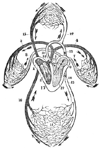
Fig.
75. An ideal view of the circulation in the lungs and system. From the right ventricle of the heart, (2,) the dark, impure blood is forced into the pulmonary artery, (3,) and its branches (4, 5) carry the blood to the left and right lung. In the capillary vessels (6, 6) of the lungs, the blood becomes pure, or of a red color, and is returned to the left auricle of the heart, (9,) by the veins, (7, 8.) From the left auricle the pure blood passes into the left ventricle, (10.) By a forcible contraction of the left ventricle of the heart, the blood is thrown into the aorta, (11.) Its branches (12, 13, 13) carry the pure blood to every organ or part of the body. The divisions and subdivisions of the aorta terminate in capillary vessels, represented by 14, 14. In these hair-like vessels the blood becomes dark colored, and is returned to the right auricle of the heart (1) by the vena cava descendens, (15,) and vena cava ascendens, (16.) The tricuspid valves (17) prevent the reflow of the blood from the right ventricle to the right auricle. The semilunar valves (18) prevent the blood passing from the pulmonary artery to the right ventricle. The mitral valves (19) prevent the reflow of blood from the left ventricle to the left auricle. The semilunar valves (20) prevent the reflow of blood from the aorta to the left ventricle.366. The muscles exercise an agency in maintaining the venous circulation at a point above what the heart could perform. As the pulsations are diminished by rest, so they are accelerated by exercise, and very much quickened by violent effort. There can be little doubt that the increased rapidity of the return of blood through the veins, is, of itself, a sufficient cause for the accelerated movements of the heart during active exercise.
Observation. The quantity of blood in different individuals varies. From twenty-five to thirty-five pounds may be considered an average estimate in a healthy adult of medium size. The time in which the blood courses through the body and returns to the heart, is different in different individuals. Many writers on physiology unconditionally limit the period to three minutes. It is undeniable that the size and health of a person, the condition of the heart, lungs, and brain, the quantity of the circulating fluid, the amount and character of the inspired air, and the amount of muscular action, exert a modifying influence. The time probably varies from three to eight minutes.
366. What causes the accelerated movements of the heart during active exercise?
Note. Let the pupil review the anatomy and physiology of the circulatory organs from fig. 75, or from anatomical outline plates, No. 6 and 7.
CHAPTER XIX.
HYGIENE OF THE CIRCULATORY ORGANS
367. If any part of the system is deprived of blood, its vitality will cease; but, if the blood is lessened in quantity to a limited extent, only the vigor and health of the part will be impaired. The following conditions, if observed, will favor the free and regular supply of blood to all portions of the system.
368. The clothing should be loosely worn. Compression of any kind impedes the passage of blood through the vessels of the compressed portion. Hence, no article of apparel should be worn so as to prevent a free flow of blood through every organ of the body.
369. The blood which passes to and from the brain, flows through the vessels of the neck. If the dressing of this part of the body is close, the circulation will be impeded, and the functions of the brain will be impaired. This remark is particularly important to scholars, public speakers, and individuals predisposed to apoplexy, and other diseases of the brain.
370. As many of the large veins lie immediately beneath the skin, through which the blood is returned from the lower extremities, if the ligatures used to retain the hose, or any other article of apparel, in proper position, be tight and inelastic, the passage of blood through these vessels will be obstructed, producing, by their distention, the varicose, or enlarged veins. Hence elastic bands should always be used for these purposes.
367–386. Give the hygiene of the circulatory organs. 367. What effect will be produced on the body if it is deprived of blood? If the blood is only lessened in quantity? 368. Why should the clothing be worn loose? 369. What is said of dressing the neck? To what persons is this remark applicable? 370. How are enlarged veins frequently produced?
371. An equal temperature of all parts of the system promotes health. A chill on one portion of the body diminishes the size of its circulating vessels, and the blood which should distend and stimulate the chilled part, will accumulate in other organs. The deficiency of blood in the chilled portion induces weakness, while the superabundance of sanguineous fluid may cause disease in another part of the system.
372. The skin should be kept not only of an equal, but at its natural temperature. If the skin is not kept warm by adequate clothing, so that chills shall not produce a contraction of the blood-vessels and a consequent paleness, the blood will recede from the surface of the body, and accumulate in the internal organs. Cleanliness of the skin is likewise necessary, for the reason, that this condition favors the free action of the cutaneous vessels.
Observation. When intending to ride in a cold day, wash the face, hands, and feet, in cold water, and rub them smartly with a coarse towel. This is far better to keep the extremities warm, than to take spirits into the stomach.
373. Exercise promotes the circulation of the blood. As the action of the muscles is one of the important agents which propel the blood through the arteries and veins, daily and regular exercise of the muscular system is required to sustain a vigorous circulation in the extremities and skin, and also to maintain a healthy condition of the system. The best stimulants to improve the sluggish circulation of an indolent patient, whose skin is pale and whose extremities are cold, are the union of vigorous muscular exercise with agreeable mental action, and the systematic application to the skin of cold water, attended with friction.
371. Why should the temperature of the body be equal? 372. Why should the skin be kept at its natural, as well as at an equal temperature? What practical observation when intending to ride in a cold day? 373. Why does exercise promote health? What are good stimulants for sluggish circulation in the indolent?
Illustration. The coach-driver and teamster throw their arms around their bodies to warm them when cold. The muscles that are called into action in swinging the arms, force a greater quantity of blood into the chilled parts, and consequently, more heat is produced.
374. When a number of muscles are called into energetic action, a greater quantity of blood will be propelled to the lungs and heart in a given time, than when the muscles are in a state of comparative inaction. It is no uncommon occurrence, that before there is a proper expansion of the respiratory organs to correspond with the frequency and energy of the movements of the muscles, there is an accumulation of blood in the lungs, attended by a painful sensation of fulness and oppression in the chest, with violent and irregular action of the heart. This condition of the organs of the chest, called congestion, may be followed by cough, inflammation of the lungs, asthma, and a structural disease of the heart.
375. To avoid these sensations and results, when we feel necessitated to walk or run a considerable distance in a short time, commence the movements in a moderate manner increasing the speed as the respiratory movements become more frequent and their expansion more extensive, so that a sufficient amount of air may be received into the lungs to purify the increased quantity of blood forced into them. The same principles should be observed when commencing labor, and in driving horses and other animals.
Observation. When a large number of muscles are called into action after repose, as when we rise from a recumbent or sitting posture, the blood is impelled to the heart with a very strong impetus. If that organ should be diseased, it may arrive there in larger quantities than can be disposed of, and death may be the result. Hence the necessity of avoiding all sudden and violent movements, on the part of those who have either a functional or structural disease of the heart.
Mention the illustration. 374. What is the effect when a number of muscles are called into energetic action? What effect has this accumulation of blood in the lungs? 375. How can such disagreeable sensations be avoided? Mention a practical observation.
376. The mind exercises no inconsiderable influence upon the circulatory organs. When an individual is stimulated by hope, or excited by anger, the heart beats more forcibly, and the arteries act more energetically, than when a person is influenced by fear, despair, or sorrow. Consequently, the system is more fully nourished, and capable of greater exertion, when the former condition obtains, than when the latter exists.
377. The quality and quantity of the blood modify the action of the heart and blood-vessels. If this fluid is abundant and pure, the circulatory vessels act with more energy than when it is deficient in quantity or defective in quality.
Illustrations. 1st. In an athletic man, whose heart beats forcibly, and whose pulse is strong, if a considerable quantity of blood is drawn from a vein, as in bleeding, the heart will beat feebly, and the pulse will become weak.
2d. When the blood is made impure by inhaling vitiated air, the action of the heart and arteries is diminished, which produces an effect similar to that which takes place when blood is drawn from a vein.
378. Hemorrhage from divided arteries should be immediately arrested. When large blood-vessels are wounded or cut, the flow of blood must be immediately stopped, or the person soon faints, and the heart ceases its action. If it is a large artery that is wounded, the blood will be thrown out in jets, or jerks, every time the pulse beats. The flow of blood can be stopped until a surgeon arrives, either by compressing the vessel between the wound and the heart, or by compressing the end of the divided artery in the wound.
376. State some of the effects that the mind has on circulation. 377. What effect have the quantity and quality of blood upon the circulatory organs? Give illustration 1st. Illustration 2d. 378. What is necessary when large blood-vessels are wounded or cut?
Fig. 76.
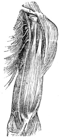
Fig. 77.
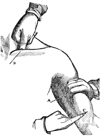
Fig. 76. The track of the large artery of the arm. 1, The collar-bone. 9, The axillary artery. 10, The brachial artery.
Fig. 77. B, The manner of compressing the artery near the collar-bone. A, The manner of compressing the large artery of the arm, with the fingers. C, The manner of compressing the divided extremity of an artery in the wound, with a finger.
379. After making compression with the fingers, as described and illustrated, take a piece of cloth or handkerchief, twist it cornerwise, and tie a hard knot midway between the two ends. This knot should be placed over the artery, between the wound and the heart, and the ends carried around the limb and loosely tied. A stick, five or six inches long, should be placed under the handkerchief, which should be twisted until the knot has made sufficient compression on the artery to allow the removal of the fingers without a return of bleeding. Continue the compression until a surgeon can be called.
What is shown by fig. 76? By fig. 77? 379. What is to be done after compressing the wound, as before described?
Fig. 78.
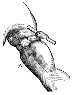
Fig. 79.
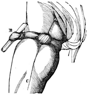
Fig. 78. A, B, The track of the large artery of the arm. The figure exhibits the method of applying the knotted handkerchief to make compression on this artery.
Fig. 79. A, C, The track of the large artery of the thigh. B, The method of applying the knotted handkerchief to compress this artery. In practice, the twisting stick B should be placed opposite the knot over the artery A, C.
380. When an artery of the arm is cut, elevating the wounded limb above the head will tend to arrest the flow of blood. In a wound of a lower limb, raise the foot, so that it shall be higher than the hip, until the bleeding ceases.
Illustration. On one occasion, the distinguished Dr. Nathan Smith was called to a person who had divided one of the large arteries below the knee. After trying in vain to find the bleeding vessel, so as to secure it, he caused the foot to be elevated higher than the hip. At the first instant the blood was forced from the wound about twelve inches; in a minute, it was diminished to three or four; and, in a short time, the bleeding ceased. This Dr. S. called his “great” operation; and it was truly great in simplicity and science.
What is shown by fig. 78 and 79? 380. What suggestion relative to the position of a limb when bleeding? Relate a simple operation by Dr. Nathan Smith.
381. The practical utility of every person knowing the proper means of arresting hemorrhage from severed arteries, is illustrated by the following incidents. In 1848, in the town of N., Mass., a mechanic divided the femoral artery; although several adult persons were present, he died in a few minutes from loss of blood, because those persons were ignorant of the method of compressing severed arteries until a surgeon could be obtained.
382. In 1846, a similar accident occurred in the suburbs of Philadelphia. While the blood was flowing copiously, a lad, who had received instruction on the treatment of such accidents at the Philadelphia High School, rushed through the crowd that surrounded the apparently dying man, placed his finger upon the divided vessel, and continued the compression until the bleeding artery was secured by a surgeon.
383. In “flesh wounds,” when no large blood-vessel is divided, wash the part with cold water, and, when bleeding has ceased, draw the incision together, and retain it with narrow strips of adhesive plaster. These should be put on smoothly, and a sufficient number applied to cover the wound. In most instances of domestic practice, the strips of adhesive plaster are too wide. They should not exceed in width one fourth of an inch. Then apply a loose bandage, and avoid all “healing salves,” ointments, and washes. In removing the dressing from a wound, both ends of the strips of plaster should be raised and drawn toward the incision. The liability of the wound re-opening is thus diminished.
381. Relate the first incident showing the utility of every person knowing the proper method of arresting the flow of blood from divided arteries. 382. The second incident.
383.
How should “flesh wounds” be dressed?Observation. The union of the divided parts is effected by the action of the divided blood-vessels, and not by salves and ointments. The only object of the dressing is to keep the parts together, and protect the wound from air and impurities. Nature, in all cases of injuries, performs her own cure. Such simple wounds do not generally require a second dressing and should not be opened until the incisions are healed.
Fig. 80.
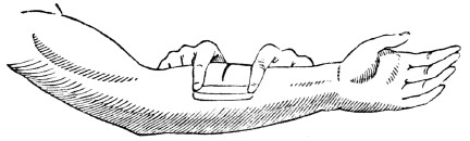
Fig. 80. The manner in which strips of adhesive plaster are applied to wounds.
384. In wounds made by pointed instruments, as a nail, or in lacerated wounds, as those made by forcing a blunt instrument, as a hook, into the soft parts, there will be no direct and immediate union. In these cases, apply a soothing poultice, as one made of linseed meal, and also keep the limb still. It is judicious to consult a physician immediately, in punctured or lacerated wounds, because they often induce the most dangerous diseases.
385. Wounds caused by the bite of rabid animals or venomous serpents, should be immediately cleansed with pure water. In many instances, the application of suction, either with “cupping glasses,” or the mouth, will prevent the introduction of the poisonous matter into the system by absorption. When this is effected, cover the wound with a soothing poultice, as one made of slippery elm bark.
What should be avoided? How should the strips of plaster be removed from a wound? How is the union of the divided parts effected? 384. How should punctured and lacerated wounds be dressed? 385. What is the treatment of wounds caused by the bite of rabid animals?
Observation. Although animal poisons, when introduced into the circulating fluid through the broken surface of the skin, frequently cause death, yet they can be taken into the mouth and stomach with impunity, if the mucous membrane which lines these parts is not broken.
Fig. 81.
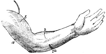
Fig. 81. a, a, Representation of wounds on the back part of the arm and fore-arm b, b, Wounds on the anterior part of the arm and fore-arm. By bending the elbow and wrist, the incisions at a, a, are opened, while those at b, b, are closed. Were the arm extended at the elbow and wrist, the wounds at a, a, would be closed, and those at b, b, would be opened.
386. The proper position of the limbs favors the union of wounds. If the incision be upon the anterior part of the leg, between the knee and ankle, extending the knee and bending the ankle will aid its closing. If it be upon the back part of the leg, by extending the foot and bending the knee, the gaping of the incision will be diminished. When wounds occur upon the trunk or upper extremities, let the position of the person be regarded.
386 Does the proper position of the limbs favor the union of wounds?
CHAPTER XX.
ABSORPTION.
387. Absorption is the process by which the materials of nutrition are removed from the alimentary canal, to be conveyed into the circulatory vessels. It is likewise the process by which the particles of matter that have become injurious, or useless, are removed from the mass of fluids and solids of which the body is composed. These renovating and removing processes are performed by two sets of vessels
ANATOMY OF THE LYMPHATIC VESSELS.
388. The vessels that act exclusively for the growth and renovation of the system, are found only in the alimentary canal. They are called lacteals. The vessels whose sole function is to remove particles of matter already deposited, are called Lym-phat´ics. The radicles, or commencement of the veins, in many, and it may be in all parts of the body, perform the office of absorption.
Observation. This fact accounts for the capacity of the venous system exceeding the arterial. Had the veins no other function to perform, beside returning the blood that had been distributed by the arteries, it would be reasonable to suppose that this system would be less than the arterial, but the reverse is known to be true.
389. The LYMPHATIC VESSELS, in structure, resemble the lacteals. They exist in great numbers in the skin and mucous membranes, particularly those of the lungs. Though no lymphatics have been traced to the brain, it is presumed that they exist there, as this part of the body is not exempt from the composition and decomposition, which are perpetual in the body. These vessels are extremely minute at their origin, so that in many parts of the system they cannot be detected without the aid of a microscope.
387. Define absorption. 388–391. Give the anatomy of the lymphatic vessels. 388. What are those vessels called that act exclusively for the growth and renovation of the body? Those whose office is to remove the atoms already deposited? What other vessels perform the office of absorption? Give observation. 389. Describe the
lymphatics.
Fig. 82.
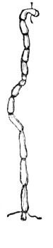
Fig. 83.
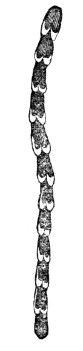
Fig. 84.
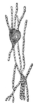
Fig. 82. A single lymphatic vessel, much magnified.
Fig. 83. The valves of a lymphatic trunk.
Fig. 84. 1, A lymphatic gland with several vessels passing through it.
390. The lymphatic vessels, like the veins, diminish in number as they increase in size, while pursuing their course toward the large veins near the heart, into which they pour their contents. The walls of these vessels have two coats of which the external one is cellular, and is capable of considerable distention. The internal coat is folded so as to form valves, like those in the veins. Their walls are so thin, that these folds give them the appearance of being knotted.
What is represented by fig. 82? By fig. 83? By fig. 84? 390. In what respect do these vessels resemble the veins of the system? Give the structure of their coats.
391. At certain points, the lymphatic vessels pass through distinct, soft bodies, peculiar to themselves, which are called lymphatic glands, which are to these vessels what the mesenteric glands are to the lacteals. The lymphatic glands vary in form and in size. They are extremely vascular, and appear to consist of a collection of minute vessels. These glands are found in different parts of the body, but are most numerous in the groins, axilla, or arm-pits, neck, and cavities of the chest and abdomen.
Observation. From exposure to cold, these glands are frequently enlarged and inflamed. They are known under the name of “kernels.” They are often diseased, particularly in scrofula, or “king’s evil.”
PHYSIOLOGY OF THE LYMPHATIC VESSELS.
392. Though the lacteals and lymphatics resemble each other in their structure and termination, yet they differ as to the nature of the fluids which they convey, as well as the nature of their functions. The lacteals open into the small intestine, and possess the power of rejecting all substances in the passing aliment, but the chyle. The lymphatics, on the contrary, not only imbibe all the various constituents of the body, both fluid and solid, but they sometimes absorb foreign and extraneous substances, when presented to their mouths, as in vaccination.
393. The varieties of absorption are, the In-ter-sti´tial, Rec-re-men-ti´tial, Ex-cre-men-ti´tial, Cu-ta´ne-ous, Res-pi´ra-to-ry, Ve´nous, and the Lac´te-al.
391. Describe the lymphatic glands. What observation is given in regard to these glands? 392–403. Give the physiology of the lymphatic vessels. 392. Explain the difference between the lacteals and lymphatics 393. Name the varieties of absorption.
394. Interstitial absorption is that change which is constantly going on in the animal economy among the particles of matter of which every texture is composed. The ordinary functions of the body, in health, require incessant action of the lymphatics; the circulatory system, with its myriads of small vessels, is constantly depositing new atoms of matter, which become vitalized, and perform a course of actions, then die, or become useless. These old atoms are removed by the absorbent system. Thus, wherever there is a minute artery to deposit a living particle of matter, there is a lymphatic vessel, or venous radicle, to remove it as soon as it shall have finished its particular office.
395. The action of the lymphatic vessels counterbalances those of nutrition, and thus the form and size of every part of the body is preserved. When their action exceeds that of the nutrient vessels, the body emaciates; when it is deficient, plethora is the result. In youth, they are less active than the nutrient vessels, and the limbs are plump; but in later periods of life, we find these actions reversed, and the body diminishes in size. It is not unfrequent that wens, and other tumors of considerable size, disappear, and even the entire bone of a limb has been removed from the same general cause. The effused fluids of bruises are also removed by absorption.
Observations. 1st. When little or no food is taken into the stomach, life is supported by the lymphatic vessels and veins imbibing the fat and reconveying it into the blood vessels. It is the removal of this secretion which causes the emaciation of the face and extremities of a person recovering from a fever. In consumption, the extreme attenuation of the limbs is caused by the absorption, not only of the fat, but also of the muscles and more solid parts of the system.
394. What is interstitial absorption? Flow are the new atoms of matter deposited? How removed? 395. What vessels do the lymphatics counterbalance in action? What is the result when their action exceeds that of the nutrient vessels? When it is less? Mention some instances of active absorption. What causes the emaciated limbs of a person recovering from fever? The extreme attenuation in consumption?
2d. Animals which live in a half torpid state during the winter, derive their nourishment from the same source. In other words, we may say the starving animal lives for a time upon itself, eating up, by internal absorption, such parts of the body as can be spared under urgent necessity, to feed those organs and continue those functions that are absolutely essential to life.
396. Recrementitial absorption is the removal of those fluids from the system, which are secreted upon surfaces that have no external outlet. These fluids are various, as the fat, the marrow, the synovia of joints, serous fluids, and the humors of the eye. Were it not for this variety of absorption, dropsy would generally exist in the cavities of the brain, chest, and abdomen, from the continued action of the secretory vessels.
397. Excrementitial absorption relates to the fluids which have been excreted, such as the bile, pancreatic fluid, saliva, milk, and other secretions.
398. Cutaneous absorption relates to the skin. Here the lymphatic vessels extend only to the cuticle, which they do not permeate. There has been much diversity of opinion on the question of cutaneous absorption; some maintaining that this membrane absorbs, while others deny it. Many experiments have proved that the skin may absorb sufficient nutriment to support life for a time, by immersing the patient in a bath of milk or broth. It has been found that the hand, immersed to the wrist in warm water, will absorb from ninety to one hundred grains of fluid in the space of an hour.
399. Thirst may be quenched by applying moist clothes to the skin, or by bathing. It is no uncommon occurrence, during a passage from one continent to the other, for the saliva to become bitter by the absorption of sea water. Medicinal substances, such as mercury, morphine, and Spanish flies, are frequently introduced into the system through the skin.
396. What is recrementitial absorption? 397. Define excrementitial absorption. 393. To what does cutaneous absorption relate? Is there a diversity of opinion respecting this variety of absorption? What do well attested experiments show? 399. What remark in reference to quenching thirst? What agency conveys medicinal substances and ointments into the system when tabbed on the skin?
400. Respiratory absorption has reference to the lungs. The mucous membrane of these organs is abundantly supplied with lymphatic vessels. By their action, substances finely pulverized, or in the form of gas, are readily imbibed when inhaled into the lungs, such as metallic vapors, odoriferous particles, tobacco smoke, and other effluvia. In this way, contagious diseases are frequently contracted.
Illustration. In inhaling sulphuric ether, or letheon, it is introduced into the vessels of the lungs in the form of vapor, and through them it is rapidly conveyed to the brain, and thus influences the nervous system.
401. Venous absorption is the function which the veins perform in absorbing from the alimentary canal liquids of various kinds that have been taken into the stomach and are not converted into chyle. In other parts of the body, they also perform the common office of lymphatics.
402. Lacteal, or digestive absorption has reference to the absorption of chyle only, which is destined for the nutrition of the body.
403. Absorption is not only very abundant, but generally very rapid, and all these varieties are maintained through life, except when suspended by disease.
400. What is said of respiratory absorption? How is letheon introduced into the system? 401. Define venous absorption. 402. What is lacteal absorption? 403. What is said of absorption?
Fig. 85.
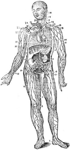
Fig. 85. A representation of the lymphatic vessels and glands. 1, 2, 3, 4, 5, 6, The lymphatic vessels and glands of the lower limbs. 7, Lymphatic glands. 8, The commencement of the thoracic duct. 9, The lymphatics of the kidney. 10, Of the stomach. 11, Of the liver. 12, 12, Of the lungs. 13, 14, 15, The lymphatics and glands of the arm. 16, 17, 18, Of the face and neck. 19, 20, Large
veins.
21, The thoracic duct. 26, The lymphatics of the heart.HYGIENE OF THE LYMPHATIC VESSELS.
404. By the action of the lymphatics, substances of an injurious, as well as of a beneficial, character may be conveyed into the system. These vessels, under certain conditions, are more active in their office than at other periods; and it is of practical utility to know what influences their action.
405. The function of these vessels is increased by moisture, and lessened by an active state of the lacteals. Observation shows that the ill-fed, and those persons that live in marshy districts, contract contagious diseases more readily than those individuals who are well fed, and breathe a dry and pure air.
406. The air of the sick-room should be dry. If due attention is not given to ventilation, the clothing of the nurse and patient, together with the air of the room, will be moistened by the exhalations from the skin and lungs. This exhalation may contain a poison of greater or less power, according to its quantity and degree of concentration, and may be absorbed and reconveyed into the system, causing inflammatory diseases, and not unfrequently death.
Observations. 1st. When we are attending a sick person a current of air that has passed over the patient should be avoided. We may approach with safety very near a person who has an infectious disease, provided care is taken to keep on the side from which the currents of air are admitted into the room.
2d. When we have been visiting or attending on a sick person, it is judicious to change the apparel worn in the sick-room, and also give the skin a thorough bathing. The outside garments, also, should be aired, as poisonous matter may have penetrated the meshes of the clothing.
404–413. Give the hygiene of the lymphatic vessels. 404. What is said respecting the action of the lymphatic vessels? 405. What influences the function of these vessels? What does observation show? 406. Why should the air of the sick-room be dry? What suggestion when we have been visiting or attending on the sick?
407. The stomach should be supplied with food of a nutrient and digestible character, in proper quantities, and at stated periods. The chyle formed from the food stimulates the lacteals to activity, which activity is attended with an inactive state of the lymphatics of the skin and lungs. Thus due attention should be given to the food of the attendants on the sick, and the members of the family. Before visiting a sick person it is judicious to take a moderate amount of nutritious food.
Observation. Many individuals, to prevent contracting disease that may be communicated from one person to another, use tobacco, either chewed or smoked; and sometimes alcohol, with decoctions of bitter herbs. These substances do not diminish, but tend to increase, the activity of the lymphatics. Thus they make use of the means by which the poisonous matter formed in the system of the diseased person, may be more readily conveyed into their
own.
408. The skin and clothing, as well as the bed-linen, should be frequently cleansed. This will remove the poisonous matter that may be deposited upon the skin and garments, which, if suffered to remain, might be conveyed into the system by the action of the lymphatics. This points also to a frequent change of the wearing apparel, as well as the coverings of the bed. In visiting the unhealthy districts of the South and West, the liability of contracting disease is much lessened by taking a supply of food at proper periods, keeping the skin and clothing in a clean state, the room well ventilated, and avoiding the damp chills of evening.
409. Absorption by the skin is most vigorous when the cuticle is removed by vesication, or blistering. Then external applications are brought into immediate contact with the orifices of the lymphatics of the skin, and by them rapidly imbibed and circulated through the system. Thus arsenic applied to the cutaneous vessels, and strong solutions of opium to extensive burns, have been absorbed in quantities sufficient to poison the patient.
407. Why should the stomach be supplied with food of a nutrient and digestible character? What is said of the use of alcohol, or tobacco, in preventing the introduction of the poisonous matter of contagious diseases? 408. Why should the clothing and bed-linen be frequently washed? What suggestion to persons in visiting the unhealthy districts of the South and West? 409. When is cutaneous absorption most vigorous? Why?
410. When the cuticle is only punctured or abraded, poisonous matter may be introduced into the system. The highly respected Dr. W., of Boston, lost his life by poisonous matter from the body of a patient subjected to a post mortem examination. He had removed from his finger, previous to the examination, a “hang-nail,” and the poison from the dead body was brought in contact with the denuded part, and through the agency of the lymphatics it was conveyed into the system.
411. Puncture any part of the cuticle with the finest instrument that has upon its point the smallest conceivable quantity of the vaccine virus, or small-pox matter, and it will be brought into contact with the lymphatic vessels, and through their agency conveyed into the system. The result is, that persons thus operated upon have the small-pox, or the vaccine disease.
412. When we expose ourselves to any poisonous vapors, or handle diseased animals or sick persons, safety and health require that the cuticle be not broken or otherwise injured. In many instances, the poisonous animal matter upon hides has been introduced into the systems of tanners, through small ulcers upon their fingers or hands. From these sores there would be seen small red lines extending up the arm. These swelled tracts indicate an inflammation of the large lymphatic trunks, that have been irritated and diseased by the passage of poisonous matter through them into the system.
410. Do the same results follow, if the cuticle is only punctured? Relate an instance of death by the absorption of poisonous matter. 411. By what means is the vaccine matter introduced into the system? 412. What caution is necessary when we expose ourselves to poisonous vapors?
Observation. A distressing illustration of the absorption of deleterious substances from the surface of a sore, is seen in the favorite experiments of that class of “quacks,” who style themselves “cancer doctors.” With them, every trifling and temporary enlargement, or tumor, is a cancer. Their
general
remedy is arsenic; and happy is the unfortunatesufferer
who escapes destruction in their hands, for toofrequently
their speedy cure is death.413. In case of an accidental wound, it is best
immediately
to bathe the part thoroughly in pure water, and to avoid all irritating applications. In some instances, it would be well to apply lunar caustic immediately. When handling or shrouding dead bodies, or removing the skin from animals that have died of disease, it would be well to lubricate the hands with olive-oil or lard. This affords protection to the minute portions of the skin, from which the cuticle may be removed. In all cases where there is an ulcer or sore, the part should be covered with something impervious to fluids, as court-plaster, before exposing the system to any animal, vegetable, or mineral poison.413. What direction is given when the cuticle is broken? What suggestion is given when shrouding dead bodies?
CHAPTER XXI.
SECRETION.
414. In the human body are found many fluids and solids of dissimilar appearance and character. These are produced by the action of organs, some of which are of simple structure, while others are very complicated in their arrangement. These organs are called Se-cre´to-ry.
ANATOMY OF THE SECRETORY ORGANS.
415. The SECRETORY ORGANS are the Ex-ha´lants,
Fol´li-cles
, and the Glands.416. The EXHALANTS were supposed to be terminations of arteries or capillaries. The external exhalants terminate on the skin and mucous membranes; the internal in the cellular and medullary tissues. (Appendix I.)
Fig. 86.

Fig. 86. A secretory follicle. An artery is seen, which supplies the material for its
secretion.
Follicles are also supplied with veins and organic nerves.417. The FOLLICLES are small bags, or sacs, situated in the true skin, and mucous membrane. The pores seen on the skin are the outlets of these bodies.
414. How are the fluids and solids of the body produced? 415–419. Give the anatomy of the secretory organs. 415. Name the secretory organs. 416. Describe the exhalants. What is represented by fig. 86? 417. Define follicles.
418. The GLANDS are soft, fleshy organs, and as various in their structure, as the secretions which it is their function to produce. Each gland is composed of many small lobules united in a compact mass, and each lobule communicates by a small duct with the principal outlet, or duct of the organ. Every gland is supplied with arteries, veins, lymphatics, and nerves. These, with the ducts, are arranged in a peculiar manner, and connected by cellular membrane.
419. There are two classes of glands, one for the modification of the fluids which pass through them, as the mesenteric and lymphatic glands; and the other for the secretion of fluids which are either useful in the animal economy, or require to be rejected from the body.
Fig. 87.
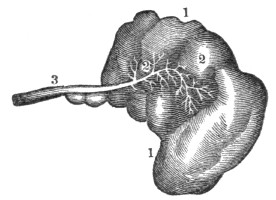
Fig. 87. 1, 1, A secretory gland. 2, 2, Minute ducts that are spread through the glands. These coalesce to form the main duct, 3.
PHYSIOLOGY OF THE SECRETORY ORGANS.
420. Secretion is one of the most obscure and
mysterious
functions of the animal economy. “It is that process by which various substances are separated from the blood, either with or without experiencing any change during their separation.” Not only is the process by which substances are separated from the blood, called secretion, but the same term is also applied to substances thus separated. Thus physiologists say, that by the process of secretion, bile is formed by the liver; and also, that bile is the secretion of this organ.418. Give the structure of the glands. 419. How are the glands arranged? 420–431. Give the physiology of the secretory organs. 420. What is
secretion
?421. The secreted fluids do not exist in that form in the blood, but most of the elements of which they are made do exist in this fluid, and the “vessels by which it is accomplished may well be called the architects and chemists of the system; for out of the same material—the blood—they construct a variety of wonderful fabrics and chemical compounds. We see the same wonderful power possessed, also, by vegetables; for out of the same materials the olive prepares its oil, the cocoa-nut its milk, the cane its sugar, the poppy its narcotic, the oak its green pulpy leaves, and its dense woody fibre. All are composed of the same few, simple elements, arranged in different order and proportions.”
422. “In like manner we find the vessels, in animated bodies, capable of forming all the various textures and substances which compose the frame; the cellular tissue, the membranes, the ligaments, the cartilages, the bones, the marrow, the muscles with their tendons, the lubricating fluid of the joints, the pulp of the brain, the transparent jelly of the eye; in short, all the textures of the various organs of which the body is composed, consist of similar ultimate elements, and are manufactured from the blood.”
423. Of the agents that produce or direct the different secretions, we have no very accurate knowledge. Some have supposed this function to be mechanical, others a chemical process, but experiments prove that it is dependent on nervous influence. If the nerves are divided which are distributed to any organ, the process of secretion is suspended. It is no uncommon occurrence, that the nature of milk will be so changed from the influence of anger in the mother, as to cause vomiting, colic, and even convulsions, in the infant that swallows it. Unexpected intelligence either of a pleasant or unpleasant character, by its influence on the nervous system, will frequently destroy the appetite. Sometimes mental agitation, as fear, will cause a cold sweat to pervade the surface of the body.
421. What is said respecting secreted substances? Do vegetables possess the property of secretion? 422. From what are the various textures formed? 423. Have we accurate knowledge of the agents that produce secretion?
424. Secretions are constantly maintained, during life, from the serous membrane, by the action of the internal exhalants. The fluid which is exhaled bears some resemblance to the serum of the blood. Its use is to furnish the organs, which are surrounded by this membrane, with a proper degree of moisture, and thus enables them to move easily on each other, as those within the chest and abdomen.
425. The cellular tissue exhales a serous fluid, and when it becomes excessive in quantity, general dropsy is produced. Fat is another secretion, which is thrown out, in a fluid state, from the cellular membrane. It is deposited in little cells, and exists in the greatest abundance between the skin and the muscles. Its use seems to be, to form a cushion around the body for its protection; to furnish nutriment for the system when food cannot be taken; to supply the carbon and hydrogen necessary to sustain the generation of heat, when these articles of combustion are not otherwise furnished. The med´ul-la-ry substance, (marrow,) in the cavities of the long bones, is very much like fat.
Observation. During sickness, if there is not emaciation or absorption of this secretion, it is considered an unfavorable symptom, because it indicates a want of power in the absorbing system, which is among the last to be affected.
How is it proved that secretion depends on nervous influence? 424. What is said of the secretions from the serous membrane? 425. From what tissue is a serous fluid exhaled? What is the effect when this fluid becomes excessive in quantity? What is fat? Its use? What is marrow?
426. The mucous secretion is a transparent, viscid fluid which is secreted by those membranes that line the cavities of the body, which have an external communication, as the trachea and alimentary canal. This secretion serves to protect these parts from the influence of the air, and concurs, by means of its peculiar properties, in the performance of their functions. 427. There are two external secretions, namely, one from the skin, called perspiration, and the other from the lungs. The cutaneous exhalation, or transpiration[14] exists in two forms, called sensible perspiration (sweat) and insensible perspiration. The pulmonary exhalation is the most important and universal, and closely resembles that of the skin.
428. The follicles are found only in the skin and mucous membrane. They secrete an oily, unctuous substance, which mixes with the transpiration, and lubricates the skin. At the root of each hair there is a minute follicle, which secretes the fluid that oils the hair. The wax in the passage of the ear is secreted from these bodies.
429. All the blood distributed to the different glands is similar in composition and character; but the fluids secreted by them, vary in appearance in a remarkable degree. The office of the glands appears to be principally to form different secretions. Thus the salivary glands secrete the insipid saliva; the lachrymal glands, the saline tears; the liver, the yellow, ropy bile; and the kidneys, the acrid urine.
426. What is said relative to the mucous secretion? 427. Name the external secretions. 428. Give the office of the follicles. 429. What appears to be the principal office of the glands? 430. Mention a secretion produced in a particular emergency.
430. Some secretions are evidently produced only in particular emergencies, as is seen in the increased secretion of bony matter when a limb is broken.
431. When any substance which is not demanded for nutrition, or does not give nourishment to the system, is imbibed by the lymphatic vessels, and conveyed into the blood, it is eliminated in the secretions.
Illustration. A few years since, a poor inebriate was carried to a London hospital in a state of intoxication. He lived but a few hours. On examining his brain, nearly half a gill of fluid, strongly impregnated with gin, was found in the cavities of this organ. This was secreted from the vessels of the brain.
HYGIENE OF THE SECRETORY ORGANS.
432. Unless the secretions are regularly maintained, disease will be the ultimate result. Let the secretions from the skin be suppressed, and fever or some internal inflammation will follow. If the bile is impeded, digestion will be impaired. If any other secretion is suppressed, it will cause a derangement of the various internal organs.
Observation. Ardent spirits derange the secretions, and change the structure of the brain. This is one reason why inebriates do not generally live to advanced age.
433. The quantity of blood influences the character of the secretions. If it is lessened to any great extent, the secretions will be lessened as well as changed in character.
Illustration. When a person has lost a considerable quantity of blood, there is a sensation of thirst in the fauces, attended with a cold, pale, dry skin. When reaction comes on, the perspiration is cold, attended with nausea, and sometimes vomiting.
431. What becomes of those substances imbibed by the lymphatics that do not give nourishment to the body. 432–437. Give the hygiene of the secretory organs. 432. What effect on the system when the secretions are not regularly maintained? 433. Does the quantity of blood influence the secretions? Give an illustration.
434. The secretory organs require the stimulus of pure blood. If this fluid is vitiated, the action of the secretory organs will be more or less modified. Either the quantity will be affected or the quality will be altered.
Observation. The impurity of the blood arising from the inhalation of the vitiated air of sleeping rooms, diminishes and changes the character of the secretions of the mouth and stomach. This accounts for the thirst, coated tongue, and disagreeable taste of the mouth when impure air is breathed during sleep. The disease it induces, is indigestion or dyspepsia.
435. The amount of action modifies the condition of the secretory organs. When a secretory organ is excessively stimulated, its vigor and energy are reduced. The subsequent debility may be so great as to suppress or destroy its functional power.
Illustrations. 1st. In those sections of the country where flax is spun on a “foot-wheel,” it is not unfrequent that the spinners moisten the thread with the secretions of the mouth. This seems to operate economically for a time, but debility of the salivary organs soon follows, which incapacitates them from supplying saliva sufficient to moisten the food, producing in a short time disease of the digestive organs.
2d. The habit of continual spitting, which attends the chewing of tobacco and gums, and other substances, between meals, induces debility, not only of the salivary glands, but of the system generally.
436. One secretory organ may do the office of another. This increased action of a secretory organ may be sustained for a limited time without permanent injury, but, if long continued, a diseased action of the organ will follow. Of morbid secretions we have examples in the ossification of the valves of the heart, cancerous and other tumors.
434. What is the effect of impure blood on the secretory organs? 435. What results from stimulating excessively a secretory organ? How is this illustrated? 436. What is the effect when one secretory organ performs the office of another?
Observation. In the evenings of the warm season, a chill upon the impressible skin, that suppresses the perspiration, is frequently followed by a diarrhœa, dysentery, or cholera morbus. These can be prevented by avoiding the chill. An efficient means of relief, is immediately to restore the skin to its proper action.
437. The secretions are much influenced by the mind. How this is effected, it is difficult to explain; but many facts corroborate it. Every one has felt an increased action of the tear-glands from distressing feelings. Cheerfulness of disposition and serenity of the passions are peculiarly favorable to the proper performance of the secretory function. From this we may learn how important it is to avoid such things as distract, agitate, or harass us.
Observation. In fevers and other diseases, when the skin, mouth, and throat are dry from a suppression of the secretions, let the mind of the patient be changed from despondency to hope, and the skin and the membrane that lines the mouth and throat will exhibit a more moist condition, together with a general improvement of the vital organs of the system. Consequently, all just encouragement of the restoration to health should be given to a sick person.
Give examples of morbid secretions. What is one cause of dysentery and cholera morbus? How can these affections he relieved? 437. Show the influence of the mind on the secretions. Mention instances of its influence.
CHAPTER XXII.
NUTRITION.
438. Nutrition is the vital act by which the different parts of the body renew the materials of which they are composed. Digestion, circulation, absorption, and respiration, are but separate links in the chain of nutrition, which would be destroyed by the absence of any one of them.
439. The nutritive process is also a kind of secretion, by which particles of matter are separated from the blood and conveyed with wonderful accuracy to the appropriate textures. The function of the nutrient vessels antagonizes those of absorption: while one system is constructing, with beautiful precision, the animal frame, the other is diligently employed in pulling down this complicated structure.
440. This ever-changing state of the body is shown by giving animals colored matter, mixed with their food, which in a short time tinges their bones with the same color as the matter introduced. Let it be withdrawn, and in a few days the bones will assume their former color—evidently from the effects of absorption. The changeful state of the body is further shown by the losses to which it is subjected; by the necessity of aliment; by the emaciation which follows abstinence from food.
438–454. What remarks respecting nutrition? 438. What is nutrition? 439. What is said of the nutritive process? The function of the nutrient vessels? 440. Give a proof of the ever-changing state of the body. Give other instances illustrative of the changeful state of the body.
441. Every part of the body is subject to this continual change of material, yet it is effected with such regularity, that the size, shape, and appearance, of every organ is preserved; and after an interval of a few years, there may not remain a particle of matter which existed in the system at a former period. Notwithstanding this entire change, the personal identity is never lost.
442. Many calculations have been made to determine in what length of time the whole body is renewed. Some have supposed that it is accomplished in four years; others have fixed the period at seven years; but the time of the change is not definite, as was supposed by a genuine son of the Emerald Isle, who had been in America seven years and three months, and consequently maintained that he was a native American.
Observation. India ink, when introduced into the skin, is not removed; hence some assert that this tissue is an exception to the alternate deposition and removal of its atoms. The ink remains because its particles are too large to be absorbed, and when in the skin it is insoluble.
443. “Those animals which are most complicated in their structure, and are distinguished by the greatest variety of vital manifestations, are subject to the most rapid changes of matter. Such animals require more frequent and more abundant supplies of food; and, in proportion as they are exposed to the greater number of external impressions, will be the rapidity of this change of matter.”
444. “Animals may be situated so that they lose nothing by secretion; consequently, they will require no nutriment. Frogs have been taken from fissures in solid lime rock, which were imbedded many feet below the surface of the earth, and, on being exposed to the air, exhibited signs of life.”
441. Why is the personal identity never lost in the change of materials, which is unceasing in the system? 442. Give the opinion of physiologists respecting the time required for the renewal of the whole body. What exception to the changing state Of the different textures? 443. What animals are subject to the most rapid changes of material? 444. May animals be situated so that they require no nutriment? What is related of frogs?
445. The renovation of the bone, muscle, ligament, tendon, cartilage, fat, nerve, hair, &c., is not perfected merely by the general circulation of the fluid which is expelled from the left side of the heart, but through the agency of a system of minute vessels, which, under ordinary circumstances, cannot be seen by the eye, even when aided by the microscope; still, minute as they are, the function of these agents is necessary to the continuance of life. They are the smallest capillary vessels.
446. “As the blood goes the round of the circulation, the nutrient capillary vessels select and secrete those parts which are similar to the nature of the structure, and the other portions pass on; so that every tissue imbibes and converts to its own use the very principles which it requires for its growth; or, in other words, as the vital current approaches each organ, the particles appropriate to it feel its attractive force,—obey it,—quit the stream,—mingle with the substance of its tissue,—and are changed into its own true and proper nature.”
447. Thus, if a bone is broken, a muscle or a nerve wounded, and, if the system is in a proper state of health, the vital economy immediately sets about healing the rupture. The blood, which flows from the wounded vessels, coagulates in the incision, for the double purpose of stanching the wound, and of forming a matrix for the regeneration of the parts. Very soon, minute vessels shoot out from the living parts into
the
coagulum of the blood, and immediately commence their operations, and deposit bony matter, where it is required to unite fractured bones, and nervous substance to heal the wounded nerve, &c.445. Show how the renovation of the bones, muscles, &c., is perfected. 446. What is said of the office of the nutrient capillary vessels? 447. When a bone is fractured, by what process is it healed?
448. But the vital economy seems not to possess the power of reproducing the muscles and true skin, and therefore, when these parts are wounded, the rupture is repaired by a gelatinous substance, which gradually becomes hard, and sometimes assumes something of a fibrous appearance. It so perfectly unites the divided muscle, however, as to restore its functional power. When the cuticle is removed, it is reproduced and no scar remains; but, when the true skin is destroyed, a scar is formed.
449. It is not uncommon that the nutrient arteries have their action so much increased in some parts, as to produce preternatural growth. Sometimes the vessels whose function it is to deposit fat, are increased in action, and wens of no inferior size are formed. Again, there may be a deposition of substances unlike any known to exist in the body. Occasionally, these nutrient arteries of a part take on a new action, and not only deposit their ordinary substance, but others, which they have not heretofore secreted, but which are formed by vessels of other parts of the body. It is in this way that we account for the bony matter deposited in the valves of the heart and brain, also the chalky deposits around the finger-joints.
450. In infancy and childhood, the function of nutrition is very active; a large amount of food is taken, to supply the place of what is lost by the action of the absorbents, and also to contribute to the growth of the body. In middle age, nutrition and absorption are more equal; but in old age, the absorbents are more active than the nutrient vessels. The size, consequently, diminishes, the parts become weaker, the bones more brittle, the body bends forward, and every function exhibits marks of decay and dissolution.
451. A striking instance of active absorption in middle age was exhibited in the person of Calvin Edson, of Vermont, who was exhibited in the large towns of New England, as the “living skeleton.” In early manhood he was athletic, and weighed one hundred and sixty pounds; but the excessive action of the absorbents over the nutrient vessels, reduced his weight, in the interval of eighteen years, to sixty pounds.
448. What occurs when a muscle is divided? 449. State some of the results of an increased action of the nutrient arteries. 450. When is nutrition most active? How in middle age? How in old age? 451. Relate a striking instance of active absorption in middle age.
452. Instances, on the other hand, have occurred, of the action of the nutrient vessels exceeding, in an extreme degree, those of absorption; as in the person of a colored girl, thirteen years of age, who was exhibited in New York in the summer of 1840. She was of the height of misses at that age, but weighed five hundred pounds. Several cases are on record of persons weighing eight hundred pounds.
453. As already mentioned, the blood is the nutritive fluid of animals. When this fluid is coagulated, a thick, jelly-like mass floats in the serum, called coagulum. This coagulated mass is composed of fibrin, and red globulated matter. The color of the red globules is owing to the presence of iron, though some physiologists think it depends on an animal substance of a gelatinous character.
Observation. That portion of the serum which remains fluid after coagulation by heat has taken place, is called se-ros´i-ty. It is more abundant in the blood of old, than in that of young animals; and it forms the “red gravy” in roasted meats.
454. The blood is not necessarily red. It may be white, as in most fish. There is no animal in which the blood is equally red in all parts of the body. The ligaments, tendons, and other white tissues in man are supplied but sparingly with red blood. The fluid that supplies these tissues is whitish.
452. Of excessive nutrition in early life. 453. Describe the parts that enter into the composition of the blood. What part of the blood forms the red gravy in roasted meats? 454. Is the blood necessarily red? Of what color is the blood of the fish? What part of the human system has white blood?
HYGIENE OF NUTRITION.
455. Healthy nutrition requires pure blood. If the nutrient arteries of the bones are supplied with impure blood, they will become soft or brittle, their vitality will be impaired, and disease will be the ultimate result. The five hundred muscles receive another portion of the blood. These organs are attached to, and act upon the bones. Upon the health and contractile energy of the muscles depends the ability to labor. Give these organs of motion impure blood, which is an unhealthy stimulus, and they will become enfeebled, the step will lose its elasticity, the movement of the arm will be inefficient, and every muscle will be incapacitated to perform its usual amount of labor.
456. When the stomach, liver, and other organs subservient to the digestion of food, are supplied with impure blood, the digestive process is impaired, causing faintness and loss of appetite, also a deranged state of the intestines, and, in general, all the symptoms of dyspepsia.
457. The delicate structure of the lungs, in which the blood is or should be purified, needs the requisite amount of pure blood to give them vigor and health. When the blood is not of this character, the lungs themselves lose their tone, and, even if permitted to expand freely, have not power fully to change the impure quality of this circulating fluid.
458. The health and beauty of the skin require that the blood should be well purified; but, if the arteries of the skin receive vitiated blood, pimples and blotches appear, and the individual suffers from “humors.” Drinks, made of various kinds of herbs, as well as pills and powders, are taken for this affection. These will never have the desired effect, while the causes of impure blood exist.
455–462. Give the hygiene of nutrition. 455. What is the effect of impure blood upon the bones? On the muscles? 456. On the digestive organs? 457. On the lungs? 458. What is the effect if the vessels of the skin are supplied with vitiated blood?
459. If the nutrient arteries convey impure material to the brain, the nervous and bilious headache, confusion of ideas, loss of memory, impaired intellect, dimness of vision, and dulness of hearing, will be experienced; and in process of time, the brain becomes disorganized, and the brittle thread of life is broken.
Observations. 1st. An exertion of any organ beyond its powers, induces weakness that will disturb the nutrition of the part that is called into action; and it recovers its energy more slowly in proportion to the excess of the exertion. The function of the organ may be totally and permanently destroyed, if the exertion is extremely violent. We sometimes see palsy produced in a muscle simply by the effort to raise too great a weight. The sight is impaired, and total blindness may be produced, by exposure to light too strong or too constant. The mind may be deranged, or idiocy may follow the excess of study or the over-tasking of the brain.
2d. When the function of an organ is permanently impaired or destroyed by over-exertion, the nutrition of the part is rendered insufficient, or is entirely arrested; and then the absorbents remove it wholly or partially, as they do every thing that is no longer useful. Thus, in palsied patients, a few years after the attack, we often find scarce any trace of the palsied muscles remaining; they are reduced almost to simple cellular tissue. The condition of the calf of the leg, in a person having a club-foot, is a familiar proof of this.
460. The blood may be made impure, by the chyle being deficient in quantity or defective in quality. This state of the chyle may be produced by the food being improper in quantity or quality, or by its being taken in an improper manner, at an improper time, and when the system is not prepared for it. The remedy for impure blood produced in any of these ways is to correct the injudicious method of using food. (See Chapters XV. and XVI.)
459. How does impure blood affect the brain? What is the effect when any organ is exerted beyond its powers? What is the effect when an organ is permanently impaired? 460. How may the blood become impure?
461. The blood may also be rendered impure, by not supplying it with oxygen in the lungs, and by the carbon not being eliminated from the system through this channel. The remedy for “impurities of the blood,” produced in this manner, would be, to carefully reduce to practice the directions in the chapters on the hygiene of the respiratory organs, relative to the free movements of the ribs and diaphragm, and the proper ventilation of rooms.
462. A retention of the waste products of the skin produces impure blood. When the vessels of the skin, by which the waste, useless material is eliminated from the system, have become inactive by improper and inadequate clothing, or by a want of cleanliness, the dead, injurious atoms of matter are retained in the circulatory vessels. The only successful method of purifying the blood and restoring health when this condition exists, is to observe the directions given relative to clothing and bathing. (See Chapters XXXIII. and XXXIV.)
Observation. If the blood has become “impure,” or “loaded with humors,” (an idea generally prevalent,) it is not and cannot be “purified” by taking patent pills, powders, drops, &c. But, on the contrary, by observing the suggestions in the preceding paragraphs, the blood can be freed of its impurities, and, what is of greater importance, such “injurious humors” will be prevented.
461. Mention another means by which the blood may be made impure. How
remedied?
462. What is the effect of want of cleanliness upon the blood? What is said respecting “humors” in the blood?Fig. 88.
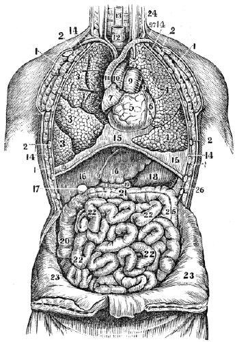
Fig. 88. A front view of the organs within the chest and abdomen. 1, 1, 1, 1, The muscles of the chest. 2, 2, 2, 2, The ribs. 3, 3, 3, The upper, middle, and lower lobes of the right lung. 4, 4, The lobes of the left lung. 5, The right ventricle of the heart. 6, The left ventricle. 7, The right auricle of the heart. 8, The left auricle. 9, The pulmonary artery. 10, The aorta. 11, The vena cava
descendens.
12, The trachea. 13, The œsophagus. 14, 14, 14, 14, The pleura. 15, 15, 15, The diaphragm. 16, 16, The right and left lobe of the liver. 17, The gall-cyst. 18, Thestomach.
26, The spleen. 19, 19, The duodenum. 20, The ascendingcolon.
21, The transverse colon. 25, The descending colon. 22, 22, 22, 22, The small intestine. 23, 23, The abdominal walls turned down. 24, The thoracic duct, opening into the left subclavian vein, (27.)CHAPTER XXIII.
THE RESPIRATORY ORGANS.
463. The nutrient portion of the food is poured into the left subclavian vein, (24, 27, fig. 88,) at the lower part of the neck, and is carried to the right cavities of the heart. The fluid in these cavities consists of the chyle incorporated with the impure blood. Neither of these two elements is fitted to promote the growth or repair the waste of the body. They must be subjected to a process, by which the first can be converted into blood, and the second freed of its carbonic acid gas and water. This is effected by the Respiratory Organs.
ANATOMY OF THE RESPIRATORY ORGANS.
464. The RESPIRATORY ORGANS are the Lungs, (lights,) the Tra´che-a, (windpipe,) the Bronch´i-a, (subdivisions of the trachea,) and the Air-Ves´i-cles, (air-cells at the extremities of the bronchia.) The Di´a-phragm, (midriff,) Ribs, and several Muscles, also aid in the respiratory process.
465. The LUNGS are conical organs, one on each side of the chest, embracing the heart, (fig. 88,) and separated from each other by a membranous partition. The color of the lungs is a pinkish gray, mottled, and variously marked with black. Each lung is divided into lobes, by a long and deep fissure, which extends from the posterior surface of the upper part of the organ, downward and forward, nearly to the anterior angle of the base. In the right lung, the upper lobe is subdivided by a second fissure. This lung is larger and shorter than the left. It has three lobes, while the left has only two.
463. What fluids are conveyed into the right cavities of the heart? What is necessary before they can be adapted to the wants of the body? By what organs are these changes effected? 464–474. Give the anatomy of the respiratory organs. 464. Name the respiratory organs. What organs also aid in the respiratory process? 465. Describe the lungs.
Fig. 89.
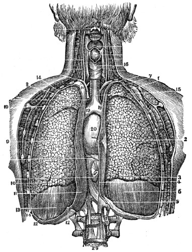
Fig. 89. A back view of the heart and lungs. The posterior walls of the chest are removed. 1, 2, 3, The upper, middle, and lower lobes of the right lung. 8, 9, 10, The two lobes of the left lung. 6, 13, The diaphragm. 7, 7, 14, 14, The pleura that lines the ribs. 4, 11, The pleura that lines the mediastine. 5, 12, 12, The portion of the pleura that covers the diaphragm. 15, The trachea, 16, The larynx. 19, 19, The right and left bronchia. 20, The heart. 29, The lower part of the spinal column.
Explain fig. 89.
466. Each lung is enclosed, and its structure maintained by a serous membrane, called the pleu´ra, which invests it as far as the root, and is thence reflected upon the walls of the chest. The lungs, however, are on the outside of the pleura, in the same way as the head is on the outside of a cap doubled upon itself. The reflected pleuræ in the middle of the thorax form a partition, which divides the chest into two cavities. This partition is called the me-di-as-ti´num.
Fig. 90.
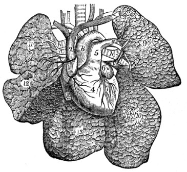
Fig. 90. The heart and lungs removed from the chest, and the lungs freed from all other attachments. 1, The right auricle of the heart. 2, The superior vena cava. 3, The inferior vena cava. 4, The right ventricle. 5, The pulmonary artery issuing from it. a, a, The pulmonary artery, (right and left,) entering the lungs. b, b, Bronchia, or air-tubes, entering the lungs. v, v, Pulmonary veins, issuing from the lungs. 6, The left auricle. 7, The left ventricle. 8, The aorta. 9, The upper lobe of the left lung. 10, Its lower lobe. 11, The upper lobe of the right
lung.
12, The middle lobe. 13, The lower lobe.Observation. When this membrane that covers the lungs, and also lines the chest, is inflamed, the disease is called “pleurisy.”
466. By what are the lungs enclosed? What is the relative position of the lungs and pleura? What is said of the reflected pleuræ? Explain fig. 90. What part of the lungs is affected in pleurisy?
467. The lungs are composed of the ramifications of the bronchial tubes, which terminate in the bronchial cells, (air-cells,) lymphatics, and the divisions of the pulmonary artery and veins. All of these are connected by cellular tissue, which constitutes the pa-ren´chy-ma. Each lung is retained in its place by its root, which is formed by the pulmonary arteries, pulmonary veins, and bronchial tubes, together with the bronchial vessels and pulmonary nerves.
468. The TRACHEA extends from the larynx, of which it is a continuation, to the third dorsal vertebra, where it divides into two parts, called bronchia. It lies anterior to the spinal column, from which it is separated by the œsophagus.
469. The BRONCHIA proceed from the bifurcation, or division of the trachea, to their corresponding lungs. Upon entering the lungs, they divide into two branches, and each branch divides and subdivides, and ultimately terminates in small sacs, or cells, of various sizes, from the twentieth to the hundredth of an inch in diameter. So numerous are these bronchial or air-cells, that the aggregate extent of their lining membrane in man has been computed to exceed a surface of 20,000 square inches, and Munro states that it is thirty times the surface of the human body.
Illustration. The trachea may be compared to the trunk of a tree; the bronchia, to two large branches; the subdivisions of the bronchia, to the branchlets and twigs; the air-cells, to the buds seen on the twigs in the spring.
470. The AIR-VESICLES and small bronchial tubes compose the largest portions of the lungs. These, when once inflated, contain air, under all circumstances, which renders their specific gravity much less than water; hence the vulgar term, lights, for these organs. The trachea and bronchial tubes are lined by mucous membrane. The structure of this membrane is such, that it will bear the presence of pure air without detriment, but not of other substances.
467. Of what are the lungs composed? How retained in place? 468. Where is the trachea situated? 469. Describe the bronchia. What is the aggregate extent of the lining membrane of the air-cells? To what may the trachea and its branches be compared? 470. What is said of the air-cells and bronchial tubes?
[14]
Transpiration is a term often used generically, to signify the passage of fluids or gases through membranes, internally or externally; but perspiration is a specific term, signifying transpiration on to the external surface.
Fig. 91.
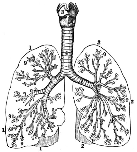
Fig. 91. A representation of the larynx, trachea, bronchia, and air-cells. 1, 1, 1, An outline of the right lung. 2, 2, 2, An outline of the left lung. 3, The larynx 4, The trachea. 5, The right bronchial tube. 6, The left bronchial tube. 7, 7, 7, 8, 8, 8, The subdivisions of the right and left bronchial tubes. 9, 9, 9, 9, 9, 9, Air-cells.
What membrane lines the trachea and its branches? What is peculiar in its structure? What does fig. 91 represent?
Observation. The structure of the trachea and lungs may be illustrated, by taking these parts of a calf or sheep and inflating the air-vesicles by forcing air into the windpipe with a pipe or quill. The internal structure may then be seen by opening the different parts.
471. The lungs, like other portions of the system, are supplied with nutrient arteries and nerves. The nervous filaments that are distributed to these organs are in part from the tenth pair, (par vagum,) that originates in the brain, and in part from the sympathetic nerve. The muscles that elevate the ribs and the diaphragm receive nervous fibres from a separate system, which is called the respiratory.
Fig. 92.
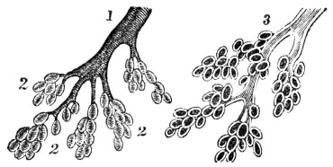
Fig. 92. 1, A bronchial tube. 2, 2, 2, Air-vesicles. Both the tube and vesicles are much magnified. 3, A bronchial tube and vesicles laid open.
Observation. When the mucous membrane of a few of the larger branches of the windpipe is slightly inflamed, it is called a “cold;” when the inflammation is greater, and extends to the lesser air-tubes, it is called bronch-i´tis. When the air-cells and parenchyma become inflamed, it is called inflammation of the lungs. Coughing is a violent expulsory effort by which air is suddenly forced through the bronchia and trachea to remove offending matter.
How may the structure of the trachea and its branches be illustrated? 471. Are the lungs supplied with nutrient arteries? Where are the respiratory nerves distributed? From what source do these organs derive their nervous filaments?
472. The RIBS are joined to the spinal column at their posterior extremity; and in front, they terminate in cartilages, which unite them to the sternum. They incline downward, from the spinal column to the breast-bone, and form resisting walls that assist in producing the partial vacuum necessary for inspiration.
Fig. 93.
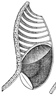
Fig. 94.
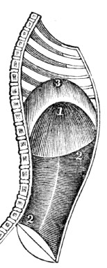
Fig. 93. A section of the chest when the lungs are inflated. 1, The diaphragm. 2, The muscular walls of the abdomen.
Fig. 94. A section of the chest when the lungs are contracted. 1, The diaphragm in common expiration. 2, 2, The muscular walls of the abdomen. 3, The position of the diaphragm in forced expiration.
These engravings show the diaphragm to be more convex, and the walls of the abdomen more flattened, when the lungs are collapsed, than when they are inflated.
473. The DIAPHRAGM is a flexible circular partition, that separates the respiratory from the digestive organs, and the chest from the abdomen. Its margin is attached to the spinal column, the sternum, and cartilages of the lower ribs. The lungs rest upon its upper surface, while the liver and stomach are placed below it, (fig. 88.) In a state of repose, its upper surface forms an arch, the convexity of which is toward the chest. In forced expiration, its upper point reaches as high as the fourth rib. In an ordinary inspiration, it is depressed as low as the seventh rib, which increases the capacity of the chest.
472. Describe the ribs. Explain figs. 93 and 94. 473. Describe the diaphragm.
474. The RESPIRATORY muscles are, in general, attached at one extremity to the parts about the shoulders, head, and upper portion of the spinal column. From these, they run downward and forward, and are attached, at the opposite extremity, to the sternum, clavicle, and upper rib. Other muscles are attached at one extremity to a rib above, and by the opposite extremity to a rib below. These fill the spaces between the ribs, and, from their situation, are called in-ter-cost´al muscles.
Observation. 1st. There are several actions of common occurrence, that are intimately connected with respiration; such as hiccough, sneezing, &c. Hiccough is an involuntary contraction of the muscles of respiration, particularly the diaphragm.
2d. Sneezing is a violent, involuntary contraction of the respiratory muscles, as in hiccough. When an acrid stimulant, as snuff, is applied to the mucous membrane of the nose, an irritation is produced which is accompanied by a violent expulsion of air from the lungs. This is owing to the connection between the nasal and respiratory nerves.
What is its form when not in action? 474. Where do the respiratory muscles make their attachment? What name is given to those muscles that fill the places between the ribs? What is hiccough? What is sneezing?
CHAPTER XXIV.
PHYSIOLOGY OF THE RESPIRATORY ORGANS.
475. Respiration, or breathing, is that process by which air is taken into the lungs and expelled from them. The object of respiration is, 1st. To supply the system with oxygen, which is essential to the generation of animal heat; 2d. To convert the chyle into blood. This is done by the oxygen of the inspired air; 3d. To relieve the organs of the body of the principal elements (carbon and hydrogen) that compose the old and useless particles of matter. The organs of the system, as already mentioned, are principally composed of carbon, hydrogen, oxygen, and nitrogen.
476. By the action of the lymphatics and capillary veins, the old and worn-out particles are conveyed into the veins of the systemic circulation. The hydrogen, in form of watery vapor, is easily discharged in the perspiration and other secretions. The nitrogen and oxygen are, or may be, separated from the blood, through the agency of several different organs; but carbon does not escape so readily. It is probable that a part of the surplus carbon of the venous blood is secreted by the liver; but a far greater amount passes to the lungs, and these may be considered as special organs designed to separate this element from the venous blood.
477. An ordinary inspiration may be accomplished by the action of the diaphragm, and a slight elevation of the ribs. In full inspiration, the diaphragm is not only more depressed but the ribs are evidently elevated. To produce this effect on the ribs, two sets of muscles are called into action. Those which are attached to the upper rib, sternum, and
clavicle
, contract and elevate the lower and free extremities of the ribs. This enlarges the cavity of the chest between the spinal column and the sternum. But the lateral diameter, in consequence, is only slightly increased, because the central portion of the ribs sinks lower than their posterior extremities, or their cartilaginous attachment to the sternum.475–494. Give the physiology of the respiratory organs. 475. What is respiration? What is the principal object in breathing? 476. How are the useless atoms of matter conveyed into the veins of the systemic circulation? How may the principal elementary substances be separated from the blood? 477. How may an ordinary inspiration be accomplished?
Fig. 95.
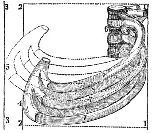
Fig. 95. 6, Four of the vertebræ, to which are attached three ribs, (7, 7, 7,) with their intercostal muscles, (8, 8.) These ribs, in their natural position, have their anterior cartilaginous extremity at 4, while the posterior extremity is attached to the vertebræ, (6,) which are neither elevated nor depressed in respiration. 1, 1, and 2, 2, parallel lines, within which the ribs lie in their natural position. If the anterior extremity of the ribs is elevated from 4 to 5, they will not lie within the line 2, 2, but will reach the line 3, 3. If two hands extend from 1, 1, to 2, 2, they will effectually prevent the elevation of the ribs from 4 to 5, as the line 2, 2, cannot be moved to 3, 3.
What effect has a full inspiration on the ribs and diaphragm? How is the chest enlarged between the spinal column and sternum? What is said of the lateral diameter of the chest? Explain fig. 95.
478. The central portion of the ribs is raised by the action of intercostal muscles. The first, or upper rib, has but little movement; the second has more motion than the first, while the third has still more than the second. The second rib is elevated by the contraction of the muscles between it and the first. The third rib is raised by the action of two sets of muscles; one lies between the first and second ribs, the other between the second and third. The motion of each succeeding rib is increased, because it is not only acted upon by the muscles that move the ribs above, but by an additional intercostal; so that the movement of the twelfth rib is very free, as it is elevated by the contraction of eleven muscles.
479. The tenth rib is raised eight times as much as the second rib, and the lateral diameter of the lower portion of the chest is increased in a corresponding degree. At the same time, the muscular margin of the diaphragm contracts, which depresses its central portion; and in this way, the chest is enlarged forward, laterally, and downward, simultaneously with the relaxation of the walls of the abdomen.
480. The lungs follow the variations of capacity in the chest, expanding their air-cells when the latter is enlarged, and contracting when the chest is diminished. Thus, when the chest is expanded, the lungs follow, and consequently a vacuum is produced in their air-cells. The air then rushes through the mouth and nose into the trachea and its branches, and fills the vacuum as fast as it is made. This mechanical process constitutes inspiration.
481. After the expansion of the chest, the muscles that elevated the ribs relax, together with the diaphragm. The elasticity of the cartilages of the ribs depresses them, and the cavity of the chest is diminished, attended by the expulsion of a portion of the air from the lungs. At the same time, the muscles that form the front walls of the abdominal cavity, contract, and press the alimentary canal, stomach, and liver, upward against the diaphragm; this, being relaxed, yields to the pressure, rises upward, and presses upon the lungs, which retreat before it, and another portion of air is expelled from these organs. This process is called expiration.
478. Describe the action of the intercostal muscles upon the ribs. 479. How does the elevation of the tenth rib compare with the second? What effect has this elevation upon the lateral diameter of the chest? 480. Describe the process of inspiration. 481. Describe the process by which the air is forced out of the lungs.
Fig. 96.
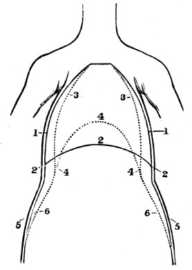
Fig. 96. A front view of the chest and abdomen in respiration. 1, 1, The position of the walls of the chest in inspiration. 2, 2, 2, The position of the diaphragm in inspiration. 3, 3, The position of the walls of the chest in expiration. 4, 4, 4, The position of the diaphragm in expiration. 5, 5, The position of the walls of the abdomen in inspiration. 6, 6, The position of the abdominal walls in
expiration.
482. Thus it is obvious that the enlargement of the chest, or inspiration, is produced in two ways: 1st. By the depression of the convex portion of the diaphragm; 2d. By the elevation of the ribs. On the contrary, the contraction of the chest, or expiration, is produced by the depression of the ribs, and elevation of the central part of the diaphragm. These movements are successive during life, and constitute respiration.
Explain fig. 96. 482. In how many ways may the chest be enlarged, and how is it accomplished? How is the contraction of the chest effected?
Fig. 97.
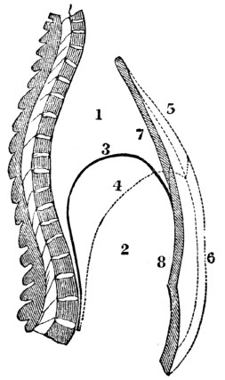
Fig. 97. A side view of the chest and abdomen in respiration. 1, The cavity of the chest. 2, The cavity of the abdomen. 3, The line of direction for the diaphragm when relaxed in expiration. 4, The line of direction for the diaphragm when contracted in inspiration. 5, 6, The position of the front walls of the chest and abdomen in inspiration. 7, 8, The position of the front walls of the abdomen and chest in expiration.
Experiment. Place the ear upon the chest of a person, and a murmuring sound will be heard, somewhat like the soft sighings of the wind through forest trees. This sound is caused by the air rushing in and out of the lungs, and is peculiarly distinct in the child.
Explain fig. 97. How may the murmur of respiration be heard?
483. It is not easy to decide how much air is taken into the lungs at each inspiration. The quantity, however, must vary in different individuals, from the difference in the condition and expansion of the lungs, together with the size of the chest. From numerous experiments, the quantity, at an ordinary inspiration, of a common-sized man, is fixed at forty cubic inches. It has been estimated that one hundred and seventy cubic inches can be thrown out of the lungs by a forcible expiration, and that there remain in the lungs two hundred and twenty cubic inches; so that these organs, in their quiescent state, may be considered as containing about three hundred and ninety cubic inches of air, or more than a gallon.
484. Respiration is more frequent in females and children than in adult men. In diseases, particularly those of the lungs, it is more increased in frequency than the action of the heart. In health, the smallest number of inspirations in a minute by an adult, is not less than fourteen, and they rarely exceed twenty-five. Eighteen may be considered an average number. The quantity of oxygen taken into the lungs at each inspiration is about eight cubic inches, one half of which disappears in every act of respiration.
Observation. Under different circumstances, however, the consumption of oxygen varies. It is greater when the temperature is low, than when it is high; and during digestion the consumption has been found one half greater than when the stomach was empty.
483. Can it be ascertained with accuracy how much air is taken into the lungs at each inspiration? Why not? What is the probable quantity that an ordinary sized man inspires? How much can be thrown out of the lungs at a forcible expiration, and how much remains in the lungs? From these calculations, how much may they contain in their quiescent state? 484. In whom is respiration most frequent? How in disease? How in health? How many may be considered an average number? When is the consumption of oxygen the greatest?
485. Dr. Southwood Smith has lately performed a series of very interesting experiments, from which he deduces the following general results: “1st. The volume of air ordinarily present in the lungs is about twelve pints. 2d. The volume of air received by the lungs at an ordinary inspiration is one pint. 3d. The volume of air expelled from the lungs at an ordinary expiration, is a little less than one pint. 4th. Of the volume of air received by the lungs at one inspiration, only one fourth part is decomposed at one action of the heart. 5th. The quantity of blood that flows to the lungs, to be acted upon by the air at one action of the heart, is two ounces, and this is acted on in less than one second of time. 6th. The quantity of blood in the whole body of the human adult, is twenty-five pounds avoirdupois, or twenty pints. 7th. In the mutual action that takes place between the air and blood, every twenty-four hours, the air loses thirty-seven ounces of oxygen, and the blood fourteen ounces of carbon.”
486. Apparently, atmospheric air is a simple element. But chemical analysis shows its composition to be oxygen and nitrogen, in the proportion of twenty-one parts of the former, and about seventy-nine of the latter. In addition, there is a small amount of vapor of water and
carbonic
acid. The pressure of this invisible, elastic fluid upon the body of an ordinary sized adult, is estimated to equal thirty-five thousand pounds.487. The principal substance of a vitiated character in the dark-colored blood is carbonic acid. And since there is no chemical affinity between the oxygen and nitrogen of the air, the former readily unites with some of the elements of the blood. Hence, whenever blood is presented to the air in the lungs, the oxygen leaves the nitrogen, and becomes mixed with the circulating fluid. (Appendix J.)
485. State the 1st, 2d, 3d, and 4th deductions from the experiments of Dr. Southwood Smith. The 5th, 6th, and 7th. 486. Of what is atmospheric air composed? What is the weight of air upon a common sized man? 487. What is the principal substance of a vitiated character in the dark-colored blood? What is said of the chemical affinity between oxygen and nitrogen?
488. Again, carbonic acid and water have a stronger affinity for atmospheric air than for the other elements of the blood. Consequently, when they are brought into contact with the air in the lungs, the carbonic acid and water leave the other constituents of the blood, and unite with the air. In this way the bluish, or impure blood is relieved of its impurities, and becomes the red, or pure blood, which contains the principles so essential to life. (Appendix K.)
489. The formation of carbonic acid and water, eliminated from the system through the lungs and skin, is explained by the following theory: In the lungs and upon the skin the oxygen separates from the nitrogen and unites with the blood in the capillary vessels of these organs. The oxygen is conveyed with the blood to the capillary arteries and veins of the different tissues of the system. In these membranes there is a chemical union of the oxygen with the carbon and hydrogen contained in the blood and waste atoms of the system. This combustion, or union of oxygen with carbon and hydrogen, is attended with the disengagement of heat, and the formation of carbonic acid and water. (Appendix L.)
490. The following experiment will illustrate the passage of fluids through membranes, and the different affinity of gases for each other. Put a mixture of water and alcohol into a phial and leave it uncorked. Both the water and alcohol have a greater affinity for air than for each other. Alcohol has a greater affinity for the air, and will be diffused through it more readily than the water, when there is no intervening obstacle. But tie a piece of bladder over the mouth of the phial, and let it stand a few days,—the water will leave the alcohol, and pass through the membrane. By the aid of this experiment, we shall endeavor to explain the interchange of fluids in the lungs.
488. What is formed when oxygen unites with carbon or hydrogen? 489. Give the theory for the formation of carbonic acid and watery vapor thrown out of the system. 490. Illustrate the passage of fluids through membranes, and the different affinities of gases.
491. The walls of the air-vesicles, and coats of the blood-vessels, are similar, in their mechanical arrangement, to the membranous bladder in the before described experiment. As the oxygen of the air has greater affinity for blood than for nitrogen, so it permeates the membranes that intervene between the air and blood more readily than the nitrogen. As the carbonic acid and water have a greater affinity for air than for the other elements of the blood, so they will also pass through the walls of the blood-vessels and air-cells more readily than the other elements of the dark-colored blood.
Fig. 98.
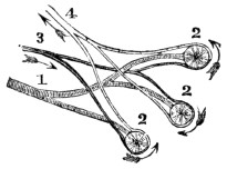
Fig. 98. 1, A bronchial tube divided into three branches. 2, 2, 2,
Air-cells.
3, Branches of the pulmonary artery, that spread over the air-cells. Through the pulmonary artery the dark, impure blood is carried to the air-cells of the lungs. 4, Branches of the pulmonary vein, that commence at the minute terminations of the pulmonary artery. Through the pulmonary vein the red blood is returned to the heart.492. As the impure blood is passing in the minute vessels over the air-cells, the oxygen passes through the thin coats of the air-cells and blood-vessels, and unites with the blood. At the same time, the carbonic acid and water leave the blood, and pass through the coats of the blood-vessels and air-cells, and mix with the air in the cells. These are thrown out of the system every time we breathe. This interchange of products produces the change in the color of the blood.
Explain fig. 98. 492. How and where is the blood changed?
Experiment. Fill a bladder with dark blood drawn from any animal. Tie the bladder closely, and suspend it in the air. In a few hours, the blood next
to
the membrane will have become of a bright red color. This is owing to the oxygen from the air passing through the bladder, and uniting with the blood, while the carbonic acid has escaped through the membrane.Fig. 99.
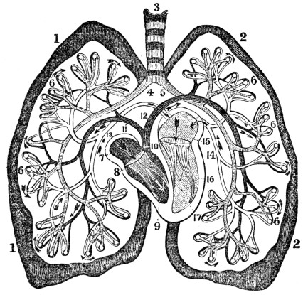
Fig. 99. An ideal view of the pulmonary circulation. 1, 1, The right lung. 2, 2, The left lung. 3, The trachea. 4, The right bronchial tube. 5, The left bronchial tube. 6, 6, 6, 6, Air-cells. 7, The right auricle. 8, The right ventricle. 9, The tricuspid valves. 10, The pulmonary artery. 11, The branch to the right lung. 12, The branch to the left lung. 13, The right pulmonary vein. 14, The left pulmonary vein. 15, The left auricle. 16, The left ventricle. 17, The mitral valves.
493. The presence of carbonic acid and watery vapor in the expired air, can be proved by the following experiments: 1st. Breathe into lime-water, and in a few minutes it will become of a milk-white color. This is owing to the carbonic acid of the breath uniting with the lime, forming the carbonate of lime. 2d. Breathe upon a cold, dry mirror for a few minutes, and it will be covered with moisture. This is condensed vapor from the lungs. In warm weather, this watery vapor is invisible in the expired air, but in a cold, dry morning in winter, the successive jets of vapor issuing from the mouth and nose are sufficiently obvious.
Give the experiment showing that oxygen changes the dark-colored blood to a bright red color. What is represented by fig. 99? 493. How can the presence of carbonic acid in the lungs be proved?
494. From the lungs are eliminated other impurities beside carbonic acid, the perceptible quality of which is various in different persons. The offensive breath of many persons may be caused by decayed teeth, or the particles of food that may be retained between them, but it often proceeds from the secretion, in the lungs, of certain substances which previously existed in the system.
Illustration. When spirituous liquors are taken into the stomach, they are absorbed by the veins and mixed with the dark-colored blood, in which they are carried to the lungs to be expelled from the body. This will explain the fact, which is familiar to most persons, that the odor of different substances is perceptible in the breath, or expired air, long after the mouth is free from these substances.
How the watery vapor? 494. Are there other excretions from the lungs? Give the illustration.
Note. Let the anatomy and physiology of the respiratory organs be reviewed from figs. 96, 97, and 99, or from anatomical outline plates Nos. 5 and
7.
CHAPTER XXV.
HYGIENE OF THE RESPIRATORY ORGANS.
495. For man to enjoy the highest degree of health, it is necessary that the impure “venous” blood be properly changed. As this is effected in the lungs by the action of the air, it follows that this element, when breathed, should be pure, or contain twenty-one per cent. of oxygen to about seventy-nine per cent. of nitrogen.
496. The volume of air expelled from the lungs is somewhat less than that which is inspired. The amount of loss varies under different circumstances. An eightieth part of the volume taken into the lungs, or half a cubic inch, may be considered an average estimate.
497. The quality and purity of the air is affected by every respiration. 1st. The quantity of oxygen is diminished. 2d. The amount of carbonic acid is increased. 3d. A certain proportion of watery vapor is ejected from the lungs in the expired air. Of the twenty-one parts of oxygen in the inspired air, only eighteen parts are expired, while the carbonic acid and watery vapor are increased about four per cent. The quantity of nitrogen is nearly the same in the expired as in the inspired air.
Observation. It is now fully ascertained that while the chemical composition of the blood is essentially changed, its weight remains the same, as the carbon and hydrogen discharged are equal to the united weight of the oxygen and nitrogen absorbed.
495–546. Give the hygiene of the respiratory organs. 495. What is necessary that man enjoy the highest degree of health? 496. How does the volume Of expired air compare with that which was inspired? Does this loss vary, and what is an average estimate? 497. How is the purity of the air affected by respiration? How is the inhaled oxygen affected? What effect on the carbonic acid and watery vapor? On the nitrogen? What is said respecting the weight of the blood?
498. If one fourth part of the volume of air received by the lungs at one inspiration is decomposed at one “beat” of the heart, it might be supposed that if the expired air be again received into the lungs, one half of the oxygen would be consumed, and, in a similar ratio, if re-breathed four times, all the oxygen would be consumed. But it does not follow, if the air is thus re-breathed, that the same changes will be effected in the lungs. For air that has been inspired does not part with its remaining oxygen as freely as when it contains the proper amount of this life-giving element, and thus the changes in the impure blood are not so completely effected.
Illustration. In the process of dyeing, each successive article immersed in the dye weakens it; but it does not follow that the dye each time is affected in the same degree, or that the coloring matter by repeated immersions can be wholly extracted. The same principle applies to the exchange of oxygen and carbonic acid gas in the lungs.
499. If the inspired air is free from moisture and carbonic acid, these substances contained in the blood will be more readily imparted to it. When the air is loaded with vapor, they are removed more slowly; but if it is saturated with moisture, no vapor will escape from the blood through the agency of the lungs. This may be illustrated by the following experiment: Take two and a half pounds of water, add to it half a pound of common salt, (chloride of sodium,) and it will readily mix with the water; and to this solution add the same quantity of salt, and it will be dissolved more slowly. Again, add more salt, and it will remain undissolved, as the water has become saturated by the pound before dissolved.
498. Does air that is re-breathed freely impart its oxygen? Why? 499. What is the effect on the blood when the air is free from vapor and carbonic acid? When loaded with vapor? When saturated? How is this illustrated?
500. The principle in this experiment is analogous to that of the union between carbonic gas and atmospheric air. Allen and Pepy showed by experiment, that air which had been once breathed, contained eight and a half per cent. of carbonic acid. They likewise showed, that no continuance of the respiration of the same air could make it take up more than ten per cent. This is the point of saturation.
Experiment. Sink a glass jar that has a stop-cock, or one with a glass stopper, into a pail of water, until the air is expelled from the jar. Fill the lungs with air, and retain it in the chest a short time, and then breathe into the jar, and instantly close the stop-cock. Close the opening of the jar that is under the water with a piece of paper laid on a plate of sufficient size to cover the opening, invert the jar, and sink into it a lighted candle. The flame will be extinguished as quickly as if put in water.[15] Remove the carbonic acid by inverting the jar, and place a lighted candle in it, and the flame will be as clear as when out of the jar.
Observations. 1st. It is familiarly known that a taper will not burn where carbonic acid exists in any considerable quantity, or when there is a marked deficiency of oxygen. From this originated the judicious practice of sinking a lighted candle into a well or pit before descending into it. If the flame is extinguished, respiration cannot there be maintained, and life would be sacrificed should a person venture in, until the noxious air is removed.
500. What did the experiments of Allen and Pepy show? How can the presence of carbonic gas in the expired air be demonstrated? State observation 1st. Observation 2d.
2d. It is the action of carbonic acid upon the respiratory organs, that gives rise to a phenomenon frequently seen in mines and caves. A man may enter these subterranean rooms, and feel no inconvenience in breathing; but the dog that follows him, falls apparently dead, and soon dies if not speedily removed to pure air. This arises from the fact that this gas is heavier than air, and sinks to the bottom of the room or cave.
3d. While it is true that carbonic acid possesses properties that render it unfit to be breathed, it is, notwithstanding, productive of very agreeable effects, when conveyed into the stomach. It forms the sparkling property of mineral waters, and fills the bubbles that rise when beer or cider is fermenting.
501. Pure atmospheric air is best adapted to a healthy action of the system. As the air cannot be maintained pure under all circumstances, the question may be asked, To what degree may the air be vitiated and still sustain life? and what is the smallest quantity of pure air a person needs each minute to maintain good health? Birnan says, that air which contains more than three and a half per cent. of carbonic acid is unfit for respiration, and, as air once respired contains eight and a half per cent. of carbonic acid, it clearly shows that it is not fitted to be breathed again.
502. No physiologist pretends that less than seven cubic feet of air are adequate for a man to breathe each minute, while Dr. Reid allows ten feet. The necessity of fifteen or twenty times the amount of air actually taken into the lungs, arises from the circumstance, that the expired air mixes with and vitiates the surrounding element that has not been inhaled.
503. The quantity of air which different persons actually need, varies. The demand is modified by the size, age, habits, and condition of the body. A person of great size who has a large quantity of blood, requires more air than a small man with a less amount of circulating fluid. Individuals whose labor is active, require more air than sedentary or idle persons, because the waste of the system is greater. On the same principle, the gormandizer needs more of this element than the person of abstemious habits. So does the growing lad require more air than an adult of the same weight, for the reason that he consumes more food than a person of mature years. Habit also exerts a controlling influence. A man who works in the open air suffers more when placed in a small, unventilated room, than one who is accustomed to breathe the confined air of workshops.
Observation 3d. 501. What questions may be asked respecting the inspired air? Give the remark of Birnan. 502. How many cubic feet of air are adequate for a man to breathe each minute? How much does Dr. Reid allow? 503. Mention some reasons why different persons do not require the same amount of
air.
504. Air, in which lamps will not burn with brilliancy, is unfitted for respiration. In crowded rooms, which are not ventilated, the air is vitiated, not only by the abstraction of oxygen and the deposition of carbonic acid, but by the excretions from the skin and lungs of the audience. The lamps, under such circumstances, emit but a feeble light. Let the oxygen gas be more and more expended, and the lamps will burn more and more feebly, until they are extinguished.
Illustrations. 1st. The effects of breathing the same air again and again, are well illustrated by an incident that occurred in one of our halls of learning. A large audience had assembled in an ill-ventilated room, to listen to a lecture; soon the lamps burned so dimly that the speaker and audience were nearly enveloped in darkness. The oppression, dizziness, and faintness experienced by many of the audience induced them to leave, and in a few minutes after, the lamps were observed to rekindle, owing to the exchange of pure air on opening the door.
How is it with the laborer? With the gormandizer? With the person that works in the open air? 504. What effect has impure air on a burning lamp? Give the illustration of the effects of impure air on lighted
lamps.
2d. In the “Black Hole of Calcutta,” one hundred and forty-six Englishmen were shut up in a room eighteen feet square, with only two small windows on the same side to admit air. On opening this dungeon, ten hours after their imprisonment, only twenty-three were alive. The others had died from breathing impure air.
505. Air that has become impure from the abstraction of oxygen, an excess of carbonic acid, or the excretions from the lungs and skin, has a deleterious effect on the body. When this element is vitiated from the preceding causes, it prevents the proper arterialization, or change in the blood. For this reason, pure air should be admitted freely and constantly into work-shops and dwelling-houses, and the vitiated air permitted to escape. This is of greater importance than the warming of these apartments. We can compensate for the deficiency of a stove, by an extra garment or an increased quantity of food; but neither garment, exercise, nor food will compensate for pure air.
506. School-rooms should be ventilated. If they are not, the pupils will be restless, and complain of languor and headache. Those unpleasant sensations are caused by a want of pure air, to give an adequate supply of oxygen to the lungs. When pupils breathe for a series of years such vitiated air, their life is undoubtedly shortened, by giving rise to consumption and other fatal diseases.
Illustration. A school-room thirty feet square and eight feet high, contains 7200 cubic feet of air. This room will seat sixty pupils, and, allowing ten cubic feet of air to each pupil per minute, all the air in the room will be vitiated in twelve minutes.
Observation. In all school-rooms where there is not adequate ventilation, it is advisable to have a recess of five or ten minutes each hour. During this time, let the pupils breathe fresh air, and open the doors and windows, so that the air of the room shall be completely changed.
Of the effects of breathing impure air. 505. In preserving health, what is of greater importance than warming the room? 506. Why should a school-room be ventilated? Give the illustration.
507. Churches, concert halls, and all rooms designed for a collection of individuals, should be amply ventilated. While the architect and workmen are assiduous in giving these public rooms architectural beauty and splendor, by adorning the ceiling with Gothic tracery, rearing richly carved columns, and providing carefully for the warming of the room, it too frequently happens that no direct provision is made for the change of that element which gives us beauty, strength, and life.
Illustration. A hall sixty feet by forty, and fifteen feet high, contains 36,000 cubic feet of air. A hall of this size will seat four hundred persons; by allowing ten cubic feet of air to each person per minute, the air of the room will be rendered unfit for respiration in nine minutes.
508. Railroad cars, cabins of steam and canal-boats, omnibuses, and stage-coaches, require ample ventilation. In the construction of these public conveyances, too frequently, the only apparent design is, to seat the greatest number of persons, regardless of the quantity and character of the air to maintain health and even life. The character of the air is only realized when, from the fresh, pure air, we enter a crowded cabin of a boat or a closed coach; then the vitiated air from animal excretions and noxious gases is offensive, and frequently produces sickness.
509. The influence of habit is strikingly expressed by Birnan, in the “Art of Warming and Ventilating Rooms:” “Not the least remarkable example of the power of habit is its reconciling us to practices which, but for its influence, would be considered noxious and disgusting. We instinctively shun approach to the dirty, the squalid, and the diseased, and use no garment that may have been worn by another. We open sewers for matters that offend the sight or the smell, and contaminate the air. We carefully remove impurities from what we eat and drink, filter turbid water, and fastidiously avoid drinking from a cup that may have been pressed to the lips of a friend. On the other hand, we resort to places of assembly, and draw into our mouths air loaded with effluvia from the lungs, skin, and clothing of every individual in the promiscuous crowd—exhalations offensive, to a certain extent, from the most healthy individuals; but when arising from a living mass of skin and lungs, in all stages of evaporation, disease, and putridity,—prevented by the walls and ceiling from escaping—they are, when thus concentrated, in the highest degree deleterious and loathsome.”
What suggestion when a school-room is not ventilated? 507. What is said in regard to ventilating churches, concert halls, &c.? State the illustration. 508. What remarks relative to public conveyances? 509. State the influence of habit by Birnan.
510. The sleeping-room should be so ventilated that the air in the morning will be as pure as when retiring to rest in the evening. Ventilation of the room would prevent morning headaches, the want of appetite, and languor—so common among the feeble. The impure air of sleeping-rooms probably causes more deaths than intemperance. Look around the country, and those who are most exposed, who live in huts but little superior to the sheds that shelter the farmer’s flocks, are found to be the most healthy and robust. Headaches, liver complaints, coughs, and a multitude of nervous affections, are almost unknown to them; not so with those who spend their days and nights in rooms in which the sashes of the windows are calked, or perchance doubled, to prevent the keen but healthy air of winter from entering their apartments. Disease and suffering are their constant companions.
510. What is said of the ventilation of sleeping-rooms? What would adequate ventilation prevent? Give a common observation.
Illustration. By many, sleeping apartments twelve feet square and seven feet high, are considered spacious for two persons, and good accommodations for four to lodge in. An apartment of this size contains 1008 cubic feet of air. Allowing ten cubic feet to each person per minute, two occupants would vitiate the air of the room in fifty minutes, and four in twenty-five minutes. When lodging-rooms are not ventilated, we would strongly recommend early rising.
511. The sick-room, particularly, should be so arranged that the impure air may escape, and pure air be constantly admitted into the room. It is no unusual practice in some communities, when a child or an adult is sick of an acute disease, to prevent the ingress of pure air, simply from the apprehension of the attendants, that the patient will contract a cold. Again, the prevalent custom of several individuals sitting in the sick-room, particularly when they remain there for several hours, tends to vitiate the air, and, consequently, to increase the suffering and danger of the sick person. In fevers or inflammatory diseases of any kind, let the patient breathe pure air; for the purer the blood, the greater the power of the system to remove disease, and the less the liability to contract colds.
Observation. Among children, convulsions, or “fits,” usually occur when they are sleeping. In many instances, these are produced by the impure air which is breathed. To prevent these alarming and distressing convulsions, the sleeping-room should be ventilated, and there should be no curtains around the bed, or coverings over the face, as they produce an effect similar to that experienced when sleeping in a small, unventilated room. To relieve a child when convulsed, carry it into the open air.
What is said of the size of sleeping-rooms? 511. What is said of the sick-room? Mention some prevailing customs in reference to these rooms. What is said of convulsions among children?
512. While occupying a room, we are insensible of the gradual vitiation of the air. This is the result of the diminished sensibility of the nervous system, and gradual adaptation of the organs to blood of a less stimulating character. This condition is well illustrated in the hibernating animals. We are insensible of the impure air of unventilated sleeping-rooms, until we leave them for a walk or ride. If they have been closed, we are made sensible of the character of the air as soon as we reënter them, for the system has regained its usual sensibility while inhaling a purer atmosphere.
513. In the construction of every inhabited room, there should be adequate means of ventilation, as well as warming. No room is well ventilated, unless as much pure air is brought into it as the occupants vitiate at every respiration. This can be effected by making an aperture in the ceiling of the room, or by constructing a ventilating flue in the chimney. This should be in contact with the flues for the escape of smoke, but separated from them by a thin brick partition. The hot air in the smoke flues will warm the separating brick partition, and consequently rarefy the air in the ventilating flue. Communication from every room in a house should be had to such flues. The draught of air can be regulated by well-adjusted registers, which in large rooms should be placed near the floor as well as near the ceiling.
514. While provision is made for the escape of rarefied impure air, we should also provide means by which pure air may be constantly admitted into the room, as the crevices of the doors and windows are not always sufficient; and, if they should be adequate, air can be introduced in a more convenient, economical, and appropriate manner. There should be an aperture opposite the ventilating flue, at or near the floor, to connect with the outer walls of the building or external
air.
But if pure heated air is introduced into the room, it obviates the necessity of the introduction of the external air.[16]512. Why are we insensible to the gradual vitiation of the air of an unventilated room? 513. What is very important in the building of every inhabited room? How can a room be well ventilated? 514. What is said relative to a communication with the external air?
515. In warming rooms, the hot air furnaces, or box and air-tight stoves converted into hot air furnaces, should be used in preference to the ordinary stoves. The air thus introduced into the room is pure as well as warm. In the adaptation of furnaces to dwelling-houses, &c., it is necessary that the air should pass over an ample surface of iron moderately heated; as a red heat abstracts the oxygen from the contiguous air, and thus renders it unfit to be respired.[17]
Observation. Domestic animals need a supply of pure air as well as man. The cows of cities, that breathe a vitiated air, have, very generally, tubercles. Sheep that are shut in a confined air, die of a disease called the “rot,” which is of a tuberculous character. Interest and humanity require that the buildings for animals be properly ventilated.
515. How should rooms be warmed? What is necessary in the adaptation of furnaces to dwelling-houses?
[15]
As a substitute for a jar with a stop-cock, take a piece of lead pipe bent in the form of a siphon, and insert it in the mouth of a reversed jar. This experiment is as conclusive whether the air is inhaled once only or breathed many times.
[17]
Dr. Wyman’s valuable work on “Ventilation,” and the work of Henry Barnard, Esq., on “School-house architecture,” can be advantageously consulted, as they give the practical methods of ventilating and warming shops, school-rooms, dwelling-houses, public halls, &c.
[16]
Mr. Frederick Emerson, of Boston, has devised a simple and effective apparatus for removing vitiated air from a room. It is successfully used upon all the public school-houses of Boston. It is now being generally applied to the school-houses and other public buildings, as well as private dwellings, of New England.